


| Received | : | Aug 25, 2026 |
| Accepted | : | Sep 17, 2026 |
| Published Online | : | Online: Sep 24, 2026 |
| Journal | : | Journal of Nanomedicine |
| Publisher | : | MedDocs Publishers LLC |
| Online edition | : | http://meddocsonline.org |
Cite this article: Ali S, Huang J. Pegylated Lipids: The Key Component in Lipid Nanoparticles for Longer Systemic Circulation. J Nanomed. 2026; 9(2): 1078.
Keywords: Lipid nanoparticles; Pegylated lipids, Systemic circulation; Efficacy; Opsonization; Novel lipids; Stealth lipids; Oncology drugs.
PEGylation first discovered for proteins to avoid immune response, has been widely recognized as a gold standard for lipid nanoparticles to prevent aggregation and reduce opsonization by blood proteins, and to avoid clearance by macrophages to help extend the longer systemic circulation [1]. The PEGylation in LNPs also helps extend stability by enhancing surface properties through steric repulsions between the particulates. The longer systemic circulation without opsonization depends upon the length of PEGylated polymeric chain and concentration around LNPs. The longer circulation helps Enhance Permeation and Retention (EPR) effect of the LNPs for drug accumulation in the cancer tissues, thereby, alleviating toxicity and increasing efficacy of the drugs [2]. Increasing concentration of PEG lipids or surface density could suppress cellular uptake and impact the endosomal escape. In addition, with repeated dosing of PEG LNPs can also trigger complement activation by inducing anti-PEG antibody production, leading to accelerated blood clearance and reducing the therapeutic efficacy of drug encapsulated in PEG-LNPs [3].
 Table 1
: Marketed drugs in LNPs.
Tenchov et al. have shown that about 65% of the approved drugs containing PEGylated lipids with oncology molecules, and other 35% used in ailments like cardiovascular, antiproliferative, anti-Alzheimer, gastrointestinal, autoimmune, neuroprotective, anti-obesity, vaccines, anti-arthritis. Over 200 clinical trials the PEGylated lipids in LNPs, have been listed on ClinicalTrials.gov. [4]. Table 1 lists the approved drugs which contain these lipids [5].
LNPs bearing the PEGylated lipids have been recognized as stealth lipids for safe and efficient delivery of a varieties of therapeutics, especially, mRNA as Covid-19 vaccines and siRNA for Onpatrro® and many others. In addition to longer circulation benefits and efficacy, PEGylated lipids can lead to unanticipated immune response by producing anti-PEG antibodies on first injection and the clearance upon subsequent injection. Other immunogenic response such as hypersensitivity reaction or Complement Activation Related Pseudo-Allergy (CARPA) also reduces safety of LNPs comprised of PEGylated lipids [6]. These are growing concerns on usage of PEGylated lipids and hence, FDA calls for safety evaluation of these long circulating lipids to assess any adverse effect by monitoring the anti-PEG antibody response of new drug candidates [7]. Reduction in cellular uptake and endosomal escape are also yet other challenges with PEGylation that require further evaluation on efficacy when selecting an excipient in drug products. Therefore, a careful assessment of the amount of PEGylation as stealth lipid to maintain its stability without interrupting drug release, is always an important consideration to overcome the PEG dilemma [8].
Structure of PEGylated lipids
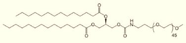
PEGylated lipids are comprised of a hydrophilic headgroup derived from phosphate tethered with polyethylene glycol varied in molecular weight with greater flexibility, and also a hydrophobic tail which is derived from long chain fatty acids, saturated or unsaturated, as shown in Figure 1. PEGylation via ester functional group is important for cleavage of PEG by enzyme in the systemic circulation. There are many other linkers through which the PEG is attached with lipids [5].
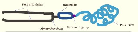
Figure 1: Structure of a typical PEGylated lipid.
Table 2
Table 2: Shows the PEGylated lipids marketed in drug products.
Let’s consider a few important biophysical attributes of the Pegylated lipids in LNPs arise from polar headgroup and negative phosphate charge, hydrophobic chains and chain lengths of the PEGs
LNP is exposed through its exterior hydrophilic surface to outer polar aqueous environment, while the hydrophobic fatty acid chains are intercalated with other fatty acid chains of the helper or phospholipid in the bilayers. These nonpolar moieties, either saturated or unsaturated, short or long, play a crucial role in bioactivities of LNPs, due, in part, by influencing the membrane curvature and fluidity, and/or by phase propensity, which is an essential requirement for regulating fusogenicity [9]. For instance, fluid membrane comprised of monounsaturated C14:1 fatty acid in LNPs, showed maximum transfection efficiency of nucleic acid complexed with a cationic phospholipid carrier [10]. The stealth properties of PEGylated lipids terminated with either methoxy or hydroxy group also showed different stealth properties. For example, those with methoxy terminal showed the best stealth with longer circulation and great systemic bioavailability as opposed to those terminating with hydroxyl group [11].
Gao’s publication sheds light on the role of PEGylated lipids in LNPs by examining the structure of PEG chains and conformations and their impact on cellular uptake and subsequently, leading to immunogenic response [14]. The polar headgroup of PEG moiety provides a hydration shield around the nanoparticles and acts as stabilizer by electrostatic interactions [15]. The higher PEG chain lengths provide greater flexibility and water solubility, therefore, it is used in a variety of drugs, in conjugation with small and large molecules, and as well as in conjugation with proteins and peptides. PEGs with MW 20-50 kD are used in oligonucleotides, siRNA, whereas, the smaller PEGs with MW 1000-5000 Da are used in larger drug formulations of antibodies and nanoparticles. The risk of potential allergic reactions such as anaphylaxis is also common with high MW PEGs such as PEG3350 and PEG5000 than PEG2000 [16]. In children, PEG3350, a known laxative, can lead to anaphylaxis [17]. LNPs comprised of PEG2000 conjugates are ideal for effective targeting with longer circulation by acting as anti-opsonization and reducing accelerated blood clearance [18].
PEGs with methoxy terminal (mPEG) vs. hydroxyl (OH) terminal possess different immunogenicity. For instance, OH-PEGs are less immunogenic than m-PEGs. This is due, in part, to lower affinity to antibodies, and hence loss of efficacy. In a study, hydroxy PEG-LNP demonstrated over 90% reduction in antibody binding with minimal complement activation compared to mPEG LNPs. Thus, OH-PEGs are preferentially used to minimize the adverse effect. In comparison with mPEG, butoxy PEGs are more immunogenic [19]. In a comparative study, PEG lipids with terminal free COOH, OH, NH2 , and MeOH, have shown that all PEGs formed the same particle size with an average PSD of 100 nm, but all PEG-lipids induces anti-PEG immunoglobin production with the exception of OH-PEG, which is being the least immunogenic and antigenic, and hence leading to rapidly clearance as compared to mPEG.
PEG surface density plays a critical parameter for performance and efficacy of PEGylated NPs. PEG’s depending upon the chain length, can arrange into mushroom or brush conformation. For example, PEG with low density, bearing short chain, can arrange in mushroom conformation while with high density and larger in chain length, can arrange in brush like conformation with extended chains. These surface densities around PEG could impact the circulation times of LNPs as well.
Hydrophobic of the PEGylated lipids, contributes to biological activity. The lengths of the fatty acid chains play a crucial role in anchoring the PEGylated lipid in the lipid membrane. The longer the chain length, stronger the hydrophobic van der Waal’s chain-chain interactions and intercalation, and vice versa. The rate of desorption of longer fatty acids during circulation is smaller compared with short chain PEGylated lipids. For instance, rate of desorption of C16 or C18 PEGylated lipids following one hour of administration was much smaller as compared to C14 PEGylated lipid in systemic circulation (45% vs. < 1.5%). The short chain fatty acids in PEG-lipids tend to dissociate into endogenous aggregates such as lipoproteins, extracellular vesicles, and blood proteins. Thus, the variations in fatty acid chain length determines the stability, and hence, the impact of LNPs on pharmacokinetics as well. PEGylated lipid comprised of C14 fatty acid can lead to dissociation from LNPs within 30 min (halflife of <0.5 h), yielding optimum hepatocyte gene silencing activity [20]. The longer fatty acid chains, like DSPE-PEG2000 while providing the stealth characteristic, lead to firm anchoring of C18 fatty acid chain to LNP bilayer with half-life of 24 h as compared to ~1.3 h half-life of a short chain DMG-PEG2000 bearing C14-fatty acid chains, leading to more effective interaction with target tissues [21].
Effects of PEGylated lipids in LNPs
Constitution of PEG lipids has direct impact on physicochemical properties of LNPs. Rabanel et al. have demonstrat ed that increasing the DMPE-PEG2000 from 1 mol% to 5% in LNPs containing C12-200, had a significant impact on particle size, Polydispersity Index (PDI), surface charge as well as on encapsulation efficiency (EE%) [22]. With increasing PEG caused the reduction in particle size from 172 nm @ 1% to 109 nm @ 5%, while the PDI increased from 0.06 to 0.146 at similar conditions, suggesting a lesser homogeneity at higher PEG levels. The surface charge on LNPs was dependent on PEG density. For instance, with increasing PEG content to 5%, the surface charge was -4.11 mV vs -2.84 mV @ 1% PEG. The encapsulation of mRNA varied within 3% and remained at 98%-58% as the PEG content increased from 1% to 5% [23]. In another study, LNPs comprised of PEGylated cholesterol with varied amounts from 1% to 5%, showed an appreciable drop in encapsulation efficiency from 90% to 80%, suggesting that drug loading dependent upon PEG density at the LNPs surface charges [24]. Taken together, it can be suggested that a rationale design approach is required to fully assess the effect of PEGylation in LNPs on particle size, charges and drug loading, and encapsulation efficiency.
PEGylation allows the LNPs to form a corona around the outer surface in aqueous phase. This hydrophilic, flexible protective layer as stealth shields the particles during systemic circulation and cellular uptake via protein adsorption. The longer PEG chain with greater steric properties is instrumental in reducing the adsorption of proteins onto LNPs surfaces. The longer or high-density PEG chain acts as brush conformation which has tendency to repel the proteins, allowing lesser affinity or binding of proteins as compared to short chain and low-density mushroom configuration. Thus, optimizing the PEGs densities with long and short chains is important to achieve the desired stealth properties without compromising the activities through surface recognition or interaction of LNPs.
Understanding the efficacy of drugs
Kumar et al. have demonstrated that LNPs encapsulating apolipoprotein (ApoE) comprised of DME-PEG2000 at 1.5%, 5% and 10% showed the identical particle size ranging 46-58 nm with stronger ApoE binding at 1.5% PEG LNP as compared to 5% PEG LNP due, in part, to reduced nonspecific binding of serum protein [25]. Intravenous administration of ApoE in LNP in C57BL/6 mice with 1.5% PEG achieved the most potent gene silencing, while LNPs with 5% and 10% PEG required much higher dosages to achieve similar results as with 1.5%. The latter results could be due to reduced adsorption of ApoE to the LNP surface. In another study, Ryals et al have demonstrated that increasing the PEG from 0.5% to 5% in DMG-PEG2000 resulted in reduction of particle size from 150 nm to 50 nm irrespective of helper lipids, DSPC and cholesterol in LNPs. On subretinal injection in BALB/c mice showed that LNPs with 0.5% PEG outperformed 5% PEG and led to 19-fold increase in luciferase expression while expressing similar ApoE levels [26].
The dosage of PEGylated lipids in LNPs remains one important consideration for balancing the efficacy and complement activation and Accelerated Blood Clearance (ABC) phenomenon. The particle size and loading all affect the ABC. On dosing in rats at >5 micromole lipid/kg, PEGylated LNPs did not show elevated anti-PEG IgM, but on dosing at < 1 micromole/ kg, ABC was considerably increased. On repeated dosing of PEGylated LNPs on day 1 and day 2 until 4 weeks, the changes in encapsulated drug clearance were not observed [27,28]. The magnitude of complement activation was at the peak by 3-4 days after the initial dosing due to expression of anti-PEG IgM. The bolus Intravenous (IV) injection of PEGylated LNPs in rats resulted in slower immune response relative to slow infusion. On the other hand, the Subcutaneous (SC) injection increased the ABC effect of PEGylated LNPs. In a study, slow IV infusion of PEGylated LNPs in pigs, caused an increase in complement activation that led to suppression of pulmonary hypertensions as compared to bolus IV injection [29]. It also led to elevation of blood anaphylatoxins levels due to complement activation. In light of these challenges, the IV infusion of Doxil® comprising of PEGylated lipids, requires slow infusion to offset the adverse effect for safe administration in the patients [30]. In fact, doxorubicin encapsulated in liposomes comprised of PEGylated lipid minimizes the production of anti-PEG IgM and prolongs the half-life of second dose. Other anticancer drugs like oxaliplatin encapsulated in PEGylated liposomes, do not induce anti-PEG IgM complement reaction [31].
Functionalized PEG lipids for targeted cellular uptake
LNPs chemically conjugated with the desired small molecules as targeting ligands help increase the cellular uptake [32]. For instance, maleimide conjugated with PEG-LNP surface is used to target cysteine residue of proteins and antibodies via thiol moieties [33]. LNPs bearing Dibenzo Cyclooctyne (DBCO) or azide group, via click chemistry undergoes azide-alkyne cycloaddition. For instance, a LNP derived from DSPE-PEG (1.5%)-maleimide undergoes conjugation with cysteine via thiol residue, which showed an enhanced mRNA transection in vitro [34]. Increasing PEG to 10% in DSPE-PEG-maleimide, for example, showed 20-fold increase in luciferase expression of ApoE in brain endothelial and neural cells, suggesting that peptide conjugation in LNPs using DSPE-PEG-maleimide retained the targeting ability. As a result, the intravenous injection dosed at 0.3 mg/kg in mice showed an enhanced efficacy with the functionalized LNPs.
Lotter et al. used dual targeted LPNs derived from (i) click chemistry by coupling of DSPE-PEG-folate with folate receptor on the breast cancer cells and (ii) coupling via cysteine thiolate of herceptin antibody ligand with DSPE-PEG-maleimide LNPs in 1:4 molar ratio [35]. The dual targeted LNPs showed a significant increase in intro activities against breast cancer cell lines including MCF7, MDA-mAb453 and SKBR3. The transfection efficiency in Her-2 overexpressing SKBR3 was 75% which was far superior over single targeting LNPs bearing folate or Herceptin receptor only, further suggesting that LNPs with multiple targeting ligands through functionalized PEG lipids can significantly enhance tumor uptake and overcome tumor heterogeneity at large.
LNPs with targeting peptides (Pep-LNPs) derived by click chemistry of DBCO on LNPs and azide on peptide were used at tumor selective mRNA delivery [36]. The resulting DSPEPEG2000-peptide conjugate was incorporated at 0.3% into LNPs with over 85% surface coverage of particles. In vitro data showed internalization or cellular uptake by PD-L1 expressing cancer cells including CT26.CL25, HCC1937 and 4T1-Luc. Intravenous injection dosing of Pep-LNPs showed 4-fold increase in tumor accumulation and 2.6-fold higher transgene expression against LNPs lacking peptide [37]. In another study, EGFR targeted LNPs were used to selective delivery of mRNA. This was achieved by derivatizing the DBCO-labeled EGFR antibody with azide functionalized PEG lipids [38]. The resultant EGFR-LNP significantly enhanced luciferase mRNA delivery in EGFR expressing JEG-3 cell. Intravenous administration at 0.4 mg/kg of EGFRLNP in mice resulted in 2-fold increase in mRNA expression due, in part, to preferential cellular uptake.
The functionalized LNPs not only serve as anchoring the proteins at target sites but are used in retention for longer circulation time. For instance, CD47 peptide functionalized LNPs with surface density of 8.9 peptide per nanoparticle at the exterior surface can increase 5-fold increase in blood retention with pronounced decrease in liver and spleen accumulation [39]. Other lipid nanoparticles modified with tetrazine conjugation with DOPE-PEG have also shown a significant potential in targeted LNP delivery [40]. Taken collectively, modified LNPs can help identify the potential targets leading to effective delivery of therapeutics and maintaining longer circulation and stability due to higher PEG density. An alternative to PEG lipids, Polysorbate 80 (PS-80) could also be used in LNPs due, in part, to inherent composition of oleic acid hydrophobic chains as tail and polyethylene glycol as hydrophilic entities as headgroup. In a comparative study, the LNPs derived from PS 80 showed 3 to 5-fold higher expression in spleen of mRNA compared to DMG-PEG2000 LNPs after intramuscular injection [41]. PS-80 LNPs encapsulating mRNA for Covid-19 vaccine induced high anti-spike IgG titers following 5 microgram single intramuscular dose.
PEGylated NPs have been recognized as safe and efficient carriers for delivery of drugs encapsulated in liposomes and polymeric nanoparticles. In spite of a few challenges, the benefits outweigh for delivery of cytotoxic drugs requiring frequent dosing and long infusion time for patients undergoing chemotherapy. Thus, the LNP technologies continue to play an important role in serving those patients requiring treatment of known ailments and rare diseases. The PEGylated lipids varied in headgroup, fatty acid chains, and the lengths and compositions of PEGs in LNPs, will have direct impact on immuno-safety, targeting and efficacy of drugs to tumor sites in preclinical and clinical evaluation.
Ascendia’s LIPIDSOL® enabling technology can help design the most robust LNPs for efficient delivery of small and large molecules and biologics. As shown in Figure 2, designed with PEGylated lipids as stealth liposomes or as lipoplexes with PEGylated lipids and ionizable lipids or cubosomes comprised of Poloxamer 407 bearing polyethylene glycol entities exposed outside in the aqueous phase, can all lead to robust formulation for efficient delivery of molecules [42]. With the continued interest in immune toxicology of LNPs, this technology will take the center stage in development of nanomedicines in the foreseeable future.
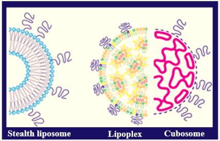
Figure 1: Illustration of PEGylated assemblies for drug delivery
We always work towards offering the best to you. For any queries, please feel free to get in touch with us. Also you may post your valuable feedback after reading our journals, ebooks and after visiting our conferences.
Table 2
| PEG-Lipid | Structure | Drug approved |
|---|---|---|
| DSPE-mPEG2000 |
 |
Doxil® |
| DMG-PEG 2000 |
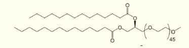 |
Moderna Spikevax®-Covid-19 Vaccine |
| ALC-0159 |
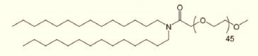 |
BioNTech/Pfizer Comirnaty® - Covid-19 vaccine |
| DMG-C-PEG2000 |
 |
Onpattro® |

