


| Received | : | Sep 17, 2026 |
| Accepted | : | Oct 02, 2026 |
| Published Online | : | Online: Oct 09 2026 |
| Journal | : | Journal of Clinical Images |
| Publisher | : | MedDocs Publishers LLC |
| Online edition | : | http://meddocsonline.org |
Cite this article: Alves da Silva FF, Bernardes ES, Passos Lima CS. Development of Graft Tumor Model of Glioblastoma in Mice. J Clin Images. 2026; 9(2): 1175.
Glioblastoma (GB) is the most aggressive brain tumor [1], with a high recurrence rate and an overall survival of only 12–14 months. Treatment involves surgical tumor resection followed by radiotherapy and/or temozolomide-based chemoradiation. However, extensive resections may cause neurological deficits that negatively affect patients quality of life, whereas incomplete resections often lead to tumor recurrence [2]. Thus, the development of in vivo models is necessary to identify more effective alternatives for the treatment and diagnosis of GB. In addition, integrins are cell-surface adhesion receptors that are overexpressed in GB [3] and have been investigated as targets for compounds labeled with positron emission tomography (PET) isotopes, such as 68Ga [4].
Therefore, this scientific communication aimed to describe the preclinical imaging obtained using an anti-integrin radiopharmaceutical with micro-PET coupled with computed tomography (μPET/CT) in a heterotopic GB graft tumor model produced with GL261 cells in female C57BL/6 mice. This study was carried out in accordance with the ARRIVE guidelines and was approved by the Ethics and Biosafety Committee of the University of Campinas (UNICAMP) under registration number CEUA/UNICAMP No. 6753-1/2025. Ten female C57BL/6 mice were used for the experiments. Each animal was injected subcutaneously on the right side of the back with 2 × 10⁶ GL261 cells in a 1:1 mixture of phosphate-buffered saline and Corning® Matrigel® Basement Membrane Matrix (catalog number 356234, Corning Life Sciences, Bedford, MA). Animals were monitored for 2–4 weeks to verify tumor growth. When the tumors reached 200 mm³, the animals were anesthetized with 2% isoflurane and positioned for imaging. For μPET/CT imaging, a 68Gaanti-RGD-integrin radiopharmaceutical (3.7–6.0 MBq) was administered intravenously to the tumor-bearing mice. Dynamic images were acquired for up to 1 hour using an Albira μPET/ CT system (Bruker Biospin Corporation, Woodbridge, CT, USA). Images were processed and quantified using PMOD software (PMOD Technologies, Zurich, Switzerland).
These representative images demonstrated that this protocol efficiently produces a heterotopic tumor-bearing in vivo model in female C57BL/6 mice, confirming successful localization of the GL261 tumor in the animals backs and supporting the use of the 68Ga-anti-RGD-integrin radiopharmaceutical as a proof of concept. Together, these results provide a reliable in vivo tumorbearing animal model for future preclinical studies aimed at developing new pharmaceutical agents against GB.
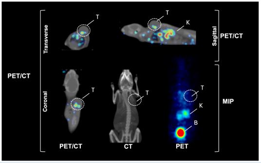
Figure 1: Non-invasive in vivo whole-body biodistribution of the 68Ga-anti-RGD-integrin radiopharmaceutical in female C57BL/6 tumorbearing mice using positron emission tomography/computed tomography (PET/CT) imaging at 1 hour. Transverse, coronal, and sagittal
profile images were obtained for imaging demonstration.
T: tumor; K: kidney; B: bladder. MIP: maximum intensity projection. Data are presented for one representative animal, identified as the
biological experimental unit (n = 10).
Acknowledgements
The study was supported by CNPq (#302922/2025-3), FAPESP (2025/26469-2), and CancerThera/CEPID (#2021/10265-8). The authors would like to thank the Radiopharmacy Center, Nuclear and Energy Research Institute (IPEN/CNEN-SP), São Paulo, Brazil.
We always work towards offering the best to you. For any queries, please feel free to get in touch with us. Also you may post your valuable feedback after reading our journals, ebooks and after visiting our conferences.

