- Case Report
- |
- Open Access
- |
- ISSN: 2637-9627
An Unique Case of Spigelian Hernia with the Complicated Appendicitis and Ipsilateral Undescended Testis in an Infant
- Dogus Guney*;
- Pediatric Surgery Clinic, Ankara Yıldırım Beyazıt University, Ankara, Turkey.
- Recep Kar;
- Pediatric Surgery Clinic, Ankara City Hospital Children’s Hospital, Ankara-Turkey.
- Betül Uysal;
- Pediatric Surgery, Ankara City Hospital Children’s Hospital, Ankara, Turkey.
- Suleyman Arif Botanci;
- Pediatric Surgery Clinic, Ankara City Hospital Children’s Hospital, Ankara-Turkey.
- Can Ihsan Oztorun;
- Pediatric Surgery Clinic, Ankara Yıldırım Beyazıt University, Ankara, Turkey.
- Ahmet Erturk;
- Pediatric Surgery Clinic, Ankara City Hospital Children’s Hospital, Ankara-Turkey.
- Sabri Demir;
- Pediatric Surgery Clinic, Ankara City Hospital Children’s Hospital, Ankara-Turkey.
- Elif Emel Erten;
- Pediatric Surgery Clinic, Ankara City Hospital Children’s Hospital, Ankara-Turkey.
- Mujdem Nur Azili;
- Pediatric Surgery Clinic, Ankara Yıldırım Beyazıt University, Ankara, Turkey.
- Emrah Senel
- Pediatric Surgery Clinic, Ankara Yıldırım Beyazıt University, Ankara, Turkey.

| Received | : | Mar 17, 2022 |
| Accepted | : | Mar 29, 2022 |
| Published Online | : | Mar 31, 2022 |
| Journal | : | Annals of Pediatrics |
| Publisher | : | MedDocs Publishers LLC |
| Online edition | : | http://meddocsonline.org |
Cite this article: Guney D, Kar R, Uysal B, Botanci SA, Oztorun CI, et al. An Unique Case of Spigelian Hernia with the Complicated Appendicitis and Ipsilateral Undescended Testis in an Infant. Ann Pediatr. 2022; 5(1): 1096.
Introduction
Lateral ventral hernia, which is also called spigelian hernia, is a rare abdominal wall defect seen in children. This hernia is caused by the herniation of preperitoneal fatty tissue and internal organs through the weakness of the aponeurosis which is between semilunar line and lateral edge of rectus muscle [1,2].
The stomach, urinary bladder, colon, appendix and other intrabadominal organs can be seen in hernia sac. In the literature, a small number of case reports has been descripted for association
of spigelian hernia and ipsilateral undescended testis; however, its etiology has not been fully elucidated [3]. The incidence of appendix in the spigelian hernia sac is very rare. Appendix in spigelian sac has been shown in total of 20 adult cases in the literature to date [4-21].
We aimed to present spigelian hernia of a 38-day-old male who had ipsilateral undescended testis and incarcerated gangrenous appendicitis in the sac as the first case in the literature in this study.
Case presentation
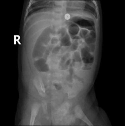
A 38-day-old boy is being evaluated with the complaint of swelling in the right lower lateral region of the abdomen and vomiting. Findings of the physical examination of patient were right spigelian hernia, an empty right hemiscrotum due to an undescended right testis and tenderness in the localization of the spigelian hernia. In addition, atypical gas distribution and partial obstruction appearance were detected in the abdominal X-ray of the patient. (Figure1).
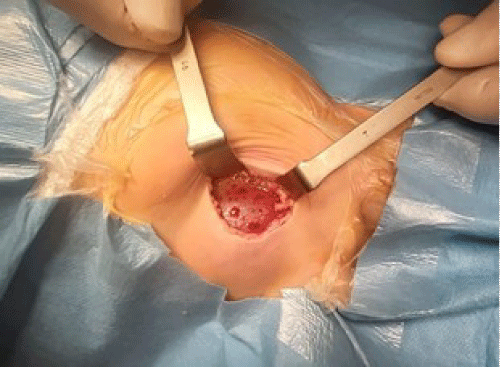
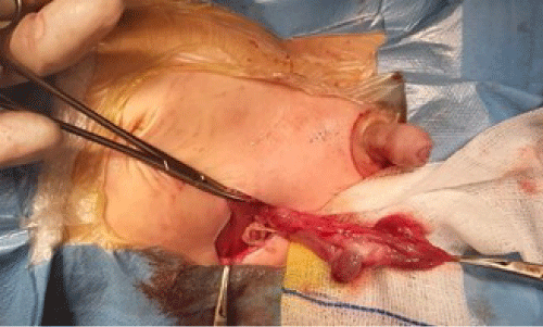
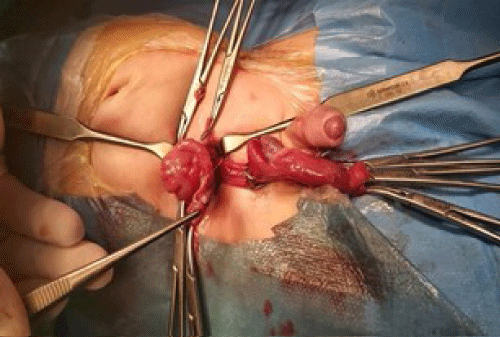
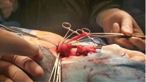
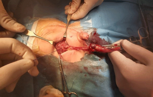
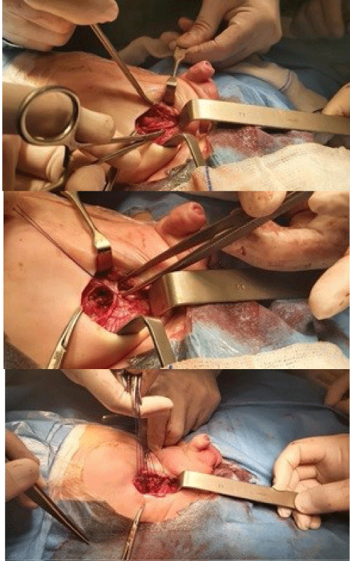
Abdominal ultrasound revealed ‘herniation of an intestinal loop with a wall thickness of 3.5 mm and testicular tissue measuring 11x9 mm from an approximately 8 mm defect in the right lower quadrant’. The patient was operated urgently (Figure 2). A right paramedian incision was made and it was seen that the right testis and asssociated structures, as well as the cecum and appendix were located in the hernia sac. Testicular appearance was normal, but spermatic cord and epididymis were edematous. The cecum appeared normal, but the appendix was gangrenous. There was no inguinal canal formation on the right. Appendectomy was performed. Orchiopexy was performed by releasing the right testis and associated structures in the retroperitoneum and descending them from the right inguinal region into the dartos pouch formed in the right hemiscrotum. Lateral ventral hernia repair was performed (Figure 3-7). No complications were observed. Patient was discharged on the post-operative third day.
Discussion
The Flemish anatomist Adriaan van der Spieghel (1578-1625) was the first to describe the semilunar line in the anatomy of the abdomen, and in 1764 Klinklosch described a hernia in the semilunar line and he defined it as a spigelian hernia[22]. In 1895, Schoofs reported the first case of pediatric spigelian hernia by presenting a case of spigelian hernia and ipsilateral undescended testis [22]. In addition, Scopinaro reported a six-month-old pediatric patient who died due to an incarcerated spigelian hernia in 1935 [3].
Spigelian hernia (lateral ventral hernia) is defined as a hernia that develops from a defect resulting from weak aponeurosis of the transverse abdominus which is between the semilunar line and the lateral edge of the rectus muscle [23]. The semilunar line starts from the ninth costal cartilage and extends to the pubic tubercle. The defect is usually not very large, most of them are less than two cm in diameter, therefore, the risk of strangulation is considered to be high. Approximately 8-25% of Spigelian hernias require immediate surgical intervention [23-25].
Spigelian hernia is more common in adults rather than children, accounting for 1-2% of all abdominal hernias. It has been shown to be associated with trauma, previous surgeries, and obesity. Spigelian hernia, which is very rare in children, is mostly congenital. It has been defined in 78 pediatric patients in the English literature search, between 1900 and 2014. 71 of these patients were reported as non-traumatic, 53% of patients had ectopic testis in the hernia sac, and 69% of cases with ectopic testis were found to have no guberneculum and/or inguinal canal [26]. In our case, there was an ectopic testis located in the spigelian sac and there was no inguinal canal on the ipsilateral side. The high prevalence of Spigelian hernia with coexisting undescended testis makes this entity a matter of debate. According to embryologhy of the testis descending, the fact that abdominal stage occurs due to intraabdominal pressure, spigelian hernia causes undescended testis. It is claimed that the testis may have remained in this region due to low resistance in the hernia localization during the abdominal descent phase [27]. Another view is that the spigelian hernia defect occurs due to undescended testis. It is argued that the testis remaining in the abdomen and absence of inguinal canal and gubernaculum structures may cause a defect in the abdominal wall, and that the coexistence of spigelian hernia and undescended testis occurs for this reason [28].
In pediatric patients testis, preperitoneal adipose tissue, small intestine, colon, omentum are frequently found in spigelian hernia sac; less frequently, Meckel's diverticulum, gallbladder, stomach, bladder, and ovaries have been reported. All of the cases in which appendix was found in the spigelian hernia sac in the literature were adult cases. In these patients, laparotomy or laparoscopic exploration, hernia repair mostly by primary or rarely patching, and appendectomy if inflamed - strangulated are recommended [4-16,29].
Pediatric spigelian hernia, ectopic testis, absence of guberneculum and absence of inguinal canal is a defined entity even though the developmental theories are still controversial. There is no reported case of incarcerated appendicitis in spigelian hernia in children. An infant patient with ipsilateral undescended testis and gangrenous appendicitis in a spigelian hernia is presented to contribute to the literature, which has no analogues in literature review
References
- Ates U, et al. Syndrome of Spigelian hernia and cryptorchidism: New evidence pertinent to pathogenic hypothesis. Urol J. 2015; 12: 2040-2041.
- Aalling L and L Penninga. Abdominal tuberculosis in a spigelian hernia. BMJ Case Rep. 2019: 12.
- Taha A, et al. Outcome of Orchidopexy in Spigelian Hernia-Undescended Testis Syndrome. Cureus. 2021; 13: e13714.
- Thomasset SC, et al. An unusual Spigelian hernia involving the appendix: a case report. Cases J. 2010; 3: 22.
- Anand R, et al. Gangrenous appendicitis contained within a Spigelian hernia. Proc (Bayl Univ Med Cent). 2020; 34: 104-106.
- Thomas MP, et al. Appendicitis in a Spigelian hernia: an unusual cause for a tender right iliac fossa mass. Ann R Coll Surg Engl. 2013; 95: e66-e68.
- Cox MJ, et al. Rare and unusual case of perforated appendicitis in a Spigelian hernia. BMJ Case Rep. 2017: 2017.
- Onal A, S Sokmen and K Atila. Spigelian hernia associated with strangulation of the small bowel and appendix. Hernia, 2003; 7: 156-157.
- Deshmukh S, et al. Computed tomographic diagnosis of appendicitis within a spigelian hernia. J Comput Assist Tomogr. 2010; 34: 199-200.
- Igwe PO and NA Ibrahim. Strangulated sliding spigelian hernia: A case report. Int J Surg Case Rep. 2018: 53: 475-478.
- Bevilacqua M, et al. Case of Spigelian hernia with incarcerated appendix. J Radiol Case Rep. 2016; 10: 23-28.
- Ndong A, et al. Strangulated spigelian hernia with necrosis of the caecum, appendix and terminal ileum: an unusual presentation in the elderly. J Surg Case Rep. 2020; 2020: 115.
- Page D and R Hendahewa. An incarcerated Spigelian hernia with the appendix in the sac passing though all layers of the abdominal wall: An unusual cause for chronic right iliac fossa pain. J Surg Case Rep. 2020; 2020: 099
- Reinke C and A Resnick. Incarcerated appendix in a Spigelian hernia. J Surg Case Rep. 2010; 2010: 3.
- Sobrado LF, et al. First case report of spigelian hernia containing the appendix after liver transplantation: Another cause for chronic abdominal pain. Int J Surg Case Rep. 2020; 72: 533-536.
- Xu L, G Dulku, and R Ho. A rare presentation of Spigelian hernia involving the appendix. Eur J Radiol Open. 2017; 4: 141-143.
- Carr JA and R Karmy-Jones. Spigelian hernia with Crohn’s appendicitis. Surg Laparosc Endosc. 1998; 8: 398-399.
- Hensley BJ and NA Stassen. Ruptured appendicitis within a left sided spigelian hernia in a patient status post previous transverse rectus abdominis myocutaneous flap resulting in necrotizing fasciitis. Am Surg. 2011; 77: E294-E295.
- Lin PH, et al. Right lower quadrant abdominal pain due to appendicitis and an incarcerated spigelian hernia. Am Surg. 2000; 66: 725-727.
- Maizlin II. Non-Operative Management of Perforated Appendicitis with in a Spigelian Hernia. Am Surg. 2021: 31348211050846.
- Ramirez-Ramirez MM and E Villanueva-Saenz. Rare hernias with atypical content: Apropos of a Spigelian hernia with acute appendicitis. Rev Gastroenterol Mex. 2017; 82: 181-182.
- Graivier L, et al. Pediatric lateral ventral (spigelian) hernias. South Med J. 988; 81: 325-326.
- Inan M, et al. Congenital Spigelian hernia associated with undescended testis. World J Pediatr. 2012; 8: 185-187.
- Polistina FA, et al. Twelve years of experience treating Spigelian hernia. Surgery. 2015; 157: 547-550.
- Spangen L. Spigelian hernia. The Surgical clinics of North America. 1984; 64: 351-366.
- Jones BC and JM Hutson. The syndrome of Spigelian hernia and cryptorchidism: A review of paediatric literature. J Pediatr Surg. 2015; 50: 325-330.
- Zekeriya ILCE RO, Mehmet Elicevik, Gonca Topuzlu Tekant. Çocuklarda nadir görülen cerrahi patoloji; lateral ventral f›t›k (Spigelian f›t›k): Olgu sunumu. Çocuk Cerrahisi Dergisi. 2007: 21: 104-107.
- Patoulias I, E Rahmani and D Patoulias. Congenital Spigelian hernia and ipsilateral cryptorchidism: A new syndrome? Folia Med Cracov. 2019; 59: 71-78.
- Spinelli C, et al. Spigelian hernia in a 14-year-old girl: A case report and review of the literature. European J Pediatr Surg Rep. 2014; 2: 58-62.


