Annals of Cardiology and Vascular Medicine
HOME /JOURNALS/Annals of Cardiology and Vascular Medicine- Clinical image
- |
- Open Access
- |
- ISSN: 2639-4383
Thrombosed Left Circumflex Artery Aneurysm Presenting with Syncope
- Sofia Lakhdar*;
- Icahn School of Medicine at Mount Sinai.
- New York City Health + Hospitals, Queens.
- Chandan Buttar;
- Icahn School of Medicine at Mount Sinai.
- New York City Health + Hospitals, Queens.
- Akwe Nyabera;
- Icahn School of Medicine at Mount Sinai.
- New York City Health + Hospitals, Queens.
- Most Sirajum Munira
- Icahn School of Medicine at Mount Sinai.
- New York City Health + Hospitals, Queens.

| Received | : | Oct 12, 2021 |
| Accepted | : | Dec 10, 2021 |
| Published Online | : | Dec 12, 2021 |
| Journal | : | Annals of Cardiology and Vascular Medicine |
| Publisher | : | MedDocs Publishers LLC |
| Online edition | : | http://meddocsonline.org |
Cite this article: Lakhdar S, Buttar C, Nyabera A, Munira MS. Thrombosed Left Circumflex Artery Aneurysmpresenting with Syncope. Ann Cardiol Vasc Med. 2021; 4(2): 1054.
Clinical image description
A 79-year-old man with history of Coronary Artery Bypass Graft (CABG), atrial fibrillation and recent abdominal aortic aneurysm repair presented after a sudden loss of consciousness. Examination revealed a blood pressure of 130/95 mm Hg, heart rate 84 beats per minute and respiratory rate 18 per minute without any distress. On physical exam, he had bilateral rales, elevated jugular venous distension, and bilateral pitting edema. Labs were significant for Troponin T increased from 0.060 to 0.120 ng/mL (Normal <=0.010 ng/mL) and Pro BNP of 3,008 pg/mL (Normal range: 1-450 pg/mL). Clinically patient’s presentation was consistent with acute decompensated heart failure.
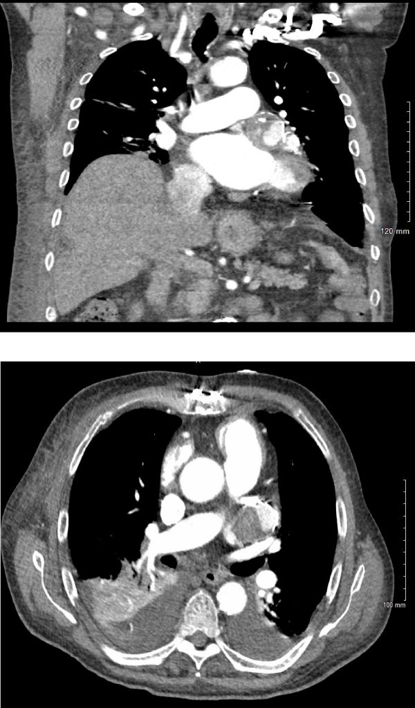
EKG was obtained showed atrial fibrillation with no specific dynamic changes. Echocardiogram revealed reduced ejection fraction and left ventricular diastolic dysfunction, bicuspid aortic valve and moderately dilated aortic root and mild dilation of the ascending aorta. Further imaging of the thoracic aorta was recommended. Contrast tomography angiography of the chest revealed coronary artery aneurysm of the left circumflex artery with mural thrombus measuring 3 cm x 3.8 cm (Figure 1). Surgical intervention was deemed risky given his overall condition. To our knowledge there has been no previously reported cases of thrombosed aneurysm in the left circumflex contributing to symptoms of heart failure or syncope as all other causes of syncope have been ruled out.
Left circumflex aneurysm thrombosis
Left circumflex artery aneurysm is an extremely rare clinical condition which requires careful evaluation of the coronary anatomy [1]. They are seen in 1.1% to 4.9% of patients undergoing coronary angiography and in about 0.02-0.04% of the general population [2]. They are commonly located in the right coronary artery. The techniques for diagnosing include noninvasive and invasive methods, such as echocardiography, CT, magnetic resonance imaging and coronary angiography. There have been no clinical trials to determine the best therapy for these patients with thrombus formation. The pathophysiology is still unclear, and the optimal treatment remains debatable. In some cases, surgical intervention is preferred. There is lack of consensus regarding the optimal management of coronary artery aneurysm; however, guideline directed medical therapy is preferred and dual antiplatelet therapy is considered if thrombosis/embolism is a concern [3].
Disclosures
The Authors report no relevant financial relationships or potential conflict of interest.
Acknowledgments
Published with the written consent of the patients.
Funding
The authors received no financial support for the research, authorship, and/or publication of this article.
Declaration of conflict of interest
The authors declare that they have no conflict of interest to report with respect to the research, authorship and/or publication of this case report.
Ethical approval
Our institution does not require ethical approval for reporting individual cases. This is an observational case report describing a patient’s clinical course. We confirm that the manuscript has been read and approved by all named authors.
Informed consent
Verbal informed consent was obtained from the patient for their anonymized information to be published in this article.
References
- Gupta V, Truong QA, Okada DR, Kiernan TJ, Yan BP, et al. Giant Left Circumflex Coronary Artery Aneurysm with Arteriovenous Fistula to the Coronary Sinus. Circulation. 2008.
- Genç B, Taştan A, Abacılar AF, Akpınar MB, Uyar S. Thrombosed left circumflex artery aneurysm presenting with myocardial infarction. Asian Cardiovasc Thorac Ann. 2016; 24: 39-41.
- Bath, Anandbir, Faizan Shaikh, Jagadeesh K. Kalavakunta. Coronary Artery Aneurysm Presenting as STEMI. BMJ Case Reports. 2019; 12: 6.
MedDocs Publishers
We always work towards offering the best to you. For any queries, please feel free to get in touch with us. Also you may post your valuable feedback after reading our journals, ebooks and after visiting our conferences.


