Annals of Cardiology and Vascular Medicine
HOME /JOURNALS/Annals of Cardiology and Vascular Medicine- Case Reports
- |
- Open Access
- |
- ISSN: 2639-4383
Plaque Modification: Need of the Hour in complex Interventional Procedures - Optical Coherence Tomography Imaging Insights
- Rama Kumari N*;
- Additional Professor of Cardiology, Department of Cardiology, Nizam’s Institute of Medical Sciences, Panjagutta, Hyderabad 500082, India
- Jeetender kumar Jain kala;
- Additional Professor of Cardiology, Department of Cardiology, Nizam’s Institute of Medical Sciences, Panjagutta, Hyderabad 500082, India
- Shabbir Ali sheik;
- Department of Cardiology, Nizam’s Institute of Medical Sciences, Panjagutta, Hyderabad 500 082, India
- Ravi Kumar reddy matli
- Department of Cardiology, Nizam’s Institute of Medical Sciences, Panjagutta, Hyderabad 500 082, India

| Received | : | Sep 18, 2020 |
| Accepted | : | Oct 20, 2020 |
| Published Online | : | Oct 22, 2020 |
| Journal | : | Annals of Cardiology and Vascular Medicine |
| Publisher | : | MedDocs Publishers LLC |
| Online edition | : | http://meddocsonline.org |
Cite this article: Rama KN, Jain kala JK, Ali sheik S, Ravi Kumar RM. Plaque Modification: Need of the Hour in complex Interventional Procedures – Optical Coherence Tomography Imaging Insights. Ann Cardiol Vasc Med. 2020: 3(1); 1030.
Keywords: Semi-compliant balloons; Non-compliant balloons; Stent expansion index; Optical coherence tomography.
Abstract
Aim: Stent under expansion is a predictor of in-stent-restenosis and stent thrombosis. Semi-Compliant Balloons (SCBs) are generally used for lesion preparation. It remains unknown whether routine pre dilatation using Non-Compliant Balloons (NCBs) improves stent expansion in calcified coronary lesions, In-stent restenosis coronary lesions.
Methods: The Pre dilatation by high-pressure NC balloon catheter for better vessel preparation and Optimal lesion preparation with non-compliant balloons for the implantation of drug eluting stent studies presenting with stable CAD or non-ST-MI requiring stent implantation to lesion preparation using NCB, SCB. Stent expansion index (which was calculated based on Optical Coherence Tomography (OCT) analysis and defined as the minimum stent/scaffold area divided by average reference lumen area (distal reference area+ proximal reference area/2).
Results: We enrolled 55 patients with calcified coronary lesions out of which 24 patients vs 31 patients divided in to NCB and SCB groups. Pre dilatation pressure was higher in the NCB group (35 ± 7 (atm) vs 14 ± 3 atm, p<0.041). Post dilatation using NCBs was performed in 24 (76%) lesions vs 31 (82%) lesions pre-treated with NCBs versus SCBs (p=0.57). Similar pressures were used for post dilatation with NCB SEB in both groups. SEI after stent implantation was in the NCB group 1.31 ± 0.30 vs 1.28 ± 0.21 in the SCB group (p=0.04). After post dilatation, SEI increased to 2.17=0.57 in the NCB group vs 2.25 ± 0.46 in the SCB group (p=0.00).
Conclusions: In calcified coronary lesions, pre-dilatation/post-dilatation with NCBs at high pressures appears to result in better scaffold and stent expansion.
Introduction
Coronary calcification is a common phenomenon in coronary artery disease. In cases of significant calcification, the coronary lesion is resistant, non-distensible, and difficult to dilate [1]. Calcified vessels frequently dissect during lesion preparation. In addition, the inability to fully dilate a lesion can lead to stent under expansion, thereby increasing the risk of restenosis and the feared complication of stent thrombosis. Lesions that cannot be dilated with a balloon catheter due to their rigidity might be amenable to cutting-balloon angioplasty [2,3]. Scoring balloons [4], rotablators [5] or the so-called “buddy wire” technique.
The use of non- compliant balloons (NCBs) for lesion preparation has become clinical routine at many sites. NCBs have a much more predictable diameter during inflation at higher pressures and the so-called ‘dog-boning’ phenomenon causing vessel dissections and perforations can be avoided. There is a lack of evidence if routine use of NCBs for lesion preparation improves stent expansion. In fact, the concept of high-pressure pre-dilatation and post-dilatation, meaning usage of inflation pressures of ≥20 atmospheres (atm), has only been propagated on the basis of case reports and case series [6-8]. Pooled analysis data of 6296 patients enrolled in seven clinical drug-eluting stents trials were analyzed to understand effects of severe coronary calcification. Patients with severe lesion calcification with 3-year follow-up showed lower rates of complete revascularization increased mortality. (10.8%) Patients without Severe Calcification (4.4%).
We assessed whether the use of OPN NC has a Rated Burst Pressure of 35 bar for cracking highly calcified lesions for lesion preparation leads to better stent expansion and is safe for percutaneous coronary interventions (PCI) in complex coronary interventions. Pre and post procedure OCT used to access the severity and degree of calcium.
Methods
Fifty -five (55) consecutive in-patients with stable CAD with calcified vessels, in-stent restenosis (ISR), or plaque preparation for calcified vessels before stent implantation, were enrolled during the period of May’2018 to Mar’2020. Ethical committee approval and informed consent from each patient were obtained. Patients with increased e GFR, acute coronary syndrome, Lesions with visible thrombus on angiogram, chronic total occlusions or bifurcation lesions were excluded.
Trial designs and Devices
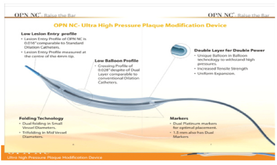
The patients were randomised by an envelope-based system. All angiograms, optical coherence tomography (OCT) recordings and clinical events were analysed in a blinded fashion by the core laboratory (KCRI). OPN NC has a Rated Burst Pressure of 35 bar for cracking highly calcified lesions. The Average Burst Pressure of OPN NC (Figure 1) is 35 bars to facilitate the plaque modification of resistant “Undilatable lesions”. After stent/scaffold implantation, OCT of the treated lesion was performed. Post dilatation was recommended in all cases of evidence for relevant stent under-expansion or malposition performed, it was mandatory to obtain a final OCT.
All patients were pre-treated with aspirin (75 mg daily dose prior to the procedure) and ticagrelor 180 mg (or in selected cases prasugrel 60 mg) before the procedure. Unfractionated heparin 70 units/kg body weight was administered intravenously at the beginning of PCI. All patients were advised to take dual antiplatelet therapy for 12 months after study enrolment.
Analysis of coronary angiography
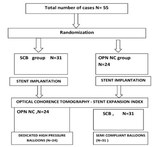
Baseline coronary target lesion characteristics and procedure results were evaluated (Figure 2). The following parameters were assessed at baseline for target lesions presence and severity of calcification, American College of Cardiology/American Heart Association (AHA) lesion severity, lesion length, reference vessel diameter, minimal luminal diameter, percentage of diameter stenosis. Calcification was defined as readily apparent densities noted within the apparent vascular wall at the stenosis and separated from the blood-filled lumen by the interceding radiolucent atheroma tissue and endothelial lining. Categories of calcification involved: none, moderate (densities noted only during the cardiac cycle prior to contrast injection) and severe (radio-opacities noted without cardiac motion prior to contrast injection generally involving both sides of the arterial wall). Of note, evaluation of target lesion after pre dilatation, stenting/scaffold-deployment and post dilatation also considered assessment of any PCI-related local complications, presence of dissections, side branch closure, distal embolization, spasm and thrombus.
Optical coherence tomography
All OCT pullbacks were also analysed off-line with dedicated software Illumine Optis, (St. Jude Medical, and USA). The analyses consisted of two main steps: (1) qualitative and quantitative evaluation of Region of Interest (ROI) and references at after-stenting pullback; (2) qualitative and quantitative evaluation of ROI and references at post dilatation pullback. Each pullback was reviewed for the presence of edge dissections, plaque protrusions, thrombus and incomplete stent apposition after the procedure.
End points and sample size justification
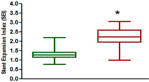
For the current analyses, our primary end point of interest was Stent Expansion Index (SEI), which was calculated based on OCT analysis and defined as the minimum stent/scaffold area divided by average reference lumen area (distal reference area + proximal reference area/2)) [9] (Figure 3). Procedural complications were systematically evaluated by using angiography and OCT. Clinical end points included new or peri-interventional Myocardial Infarction (MI) according to the guideline definitions and stent thrombosis, as defined by the academic research consortium’s definitions [10,11].
Figure 3: Stent Expansion Index (SEI) assessed by optical coherence tomography (A) after device implantation and post-dilatation in the semi-compliant balloon (SCB) group vs non-compliant balloon (NCB) group, (B) SEI after Pre-dilatation and post-dilatation in the NCB group.
Statistical analysis
The analyses were performed using SPSS V 20. Results are presented on the analyses performed according to intention-to- treat (ITT) principle, because it tends to avoid over optimistic estimates of efficacy and because of small sample size. For comparison of the baseline demographics according to allocated group’s differences in continuous variables were analysed by t-tests, Mann-Whitney U test or paired t-test as appropriate and differences in categorical variables by χ2 tests. A p value <0.05was considered statistically significant.
Results
In our study Fifty-five (55) consecutive in-patients from the Department of Cardiology were enrolled in this study during the period May 2018 to Mar 2020. The Clinical Characteristics are summarized in Table 1. The percentage of diabetics were 18.8 patients (12%), smokers were 15.4 (16%) and hypertensive were 19.3 (26.9%). Their mean age was 57.69 years. 4 had a history of previous PCI, and 1 had undergone coronary artery bypass grafting. The clinical presentation was stable angina in 4 patients, unstable angina in 2, and myocardial infarction due to graft master occultation in 1 case. Among the angiographic characteristics (Table 2), the culprit vessel was the left main coronary artery in 3 patients, the left anterior descending coronary artery was (LAD) in 40, the left circumflex coronary artery in 5, and the right coronary artery in 7. In our study 27 patients had 90 to 180 degrees’ arc of calcification on the angiogram. The OPN NC balloon was used for stent post dilation due to under expansion of graft master occlusion in one patient and same balloon used for treatment of ISR in 7 patients, intravascular ultrasonography was used in the case of plaque preparation, just in order to check for dissection, because the patient could not be stented due to the impossibility of breaking the plaque.
Procedural characteristics
Angiographic and procedural characteristics are depicted in table 2. AHA lesion classification was done for all the patients and presence and severity of angiographically visible calcification was done in all patients, as was lesion length was (22.1 ± (7.0) in OPN NC, 24.4 ± (8.3) in SEB cases p=0.07). The diameter of the balloons used for pre-dilatation was similar in both cases. Post dilatation was performed in all patients. Similar diameter sizes of post dilatation balloons were used. The diameter of the mean of balloons used for pre-dilatation were as 3.0 X 10mm. Mean pre-dilatation pressure was 35 mm Hg atm in all patients.
OCT and angiographic measurements
OCT measurements are summarised in table 3. NCB fulfilled the core-lab quality criteria for OCT assessment and were included in this analysis. Distal and proximal reference area was calculated. After stent implantation, in-stent minimal luminal diameter was slightly significantly better in the NCB group post-dilatation in-stent minimal luminal diameter increased immediately after stent implantation, SEI was 1.3 ± 0.30 in the NCB, SEI was 1.28 ± 0.21 in the SEB cases. Whereas SEI<1 4.16%, in the NCB group and 12.9% in SEB cases respectively. Patients in the after post-dilatation, SEI increased to 2.17 ± 0.59 in the NCB group and 2.25 ± 0.46 p= 0.08. Finally, the fraction of patients with SEI <0 dropped in all patients in the NCB group after post dilatation. When analysing the patients treated with NCB we found that final SEI<1 was statistically significant.
Peri- procedural safety
Peri- procedural OCT and angiographic safety outcomes are accessed. Overall, peri-procedural safety was good and no perforations of coronary artery occurred. Minor proximal and distal edge dissections, thrombus in the stented segment, side branch closure, and distal embolization were mainly seen on OCT as shown in the table 4.
Table 3: lesion assessment based on Oct measurements after stent/scaffold implantation and after Post dilatation.
Discussion
When analysing pooled data from two proof-of- concept studies including unselected patients with CAD requiring PCI, we found that the use of NCBs at high pressure for pre-dilatation and post dilatation results in better stent expansion compared with pre-dilatation with SCB. This approach appears to especially improve final SEI when implanting DES stents. Additionally, we showed that the use of high-pressure inflations with dedicated NCBs was safe and did not lead to any relevant coronary dissections or vessel perforations.
In this context, the minimum stent area reflecting stent expansion post implantation is an important independent predictor of ISR and SEI [12]. Theoretically, three measures can be considered to achieve the largest minimum stent area or SEI possible and are adequate lesion preparation, optimal stent/scaffold sizing and appropriate post dilatation once the stent/scaffold is implanted. In our study, better stent expansion was achieved immediately post device implantation when an NCB was used for lesion preparation. Post dilatation was performed with good results.
Dedicated NCBs are able to tackle the calcified and fibrotic parts of the lesion, especially if a high balloon-to- vessel ratio is used. Additionally, with NCBs increasing pressures beyond nominal values leads to an exponential rise in balloon diameter. Contrary to this, there is a linear diameter-pressure relationship in NCBs, allowing the use of higher pressure, with less risk for coronary artery rupture. For instance, the OPN NC balloon can be safely inflated above rated burst pressure of 45 atm with minimal increase in diameter and appears therefore an ideal device for complicated fibro-calcific lesions, with recoil tendency [13].
A recent post hoc analysis of two randomised studies using contemporary DES demonstrated that adjunct post dilatation was not associated with a reduction of major adverse cardiac events at 1 year among patients treated with everolimus-eluting stents, irrespective of lesion length or vessel diameter [14].
Putting the results of our study into clinical perspective, the postulated lack of efficacy of post dilatation might not be surprising since direct stenting or lesion preparation with smaller SCBs are still very popular in most catheterisation laboratories. It is obvious that many coronary lesions can probably be treated without aggressive pre-dilatation. However, interventionists should vigorously ensure optimal stent expansion and apposition in order to avoid adverse short-term and long-term outcomes, including restenosis or stent/scaffold thrombosis. As indicated by our results, achieving optimal stent expansion might require both adequate pre-dilatation and post dilatation. This concept is novel and might lead to paradigm change when addressing lesion preparation in the future.
Conclusion
OPN NC is an ultra- high pressure Plaque Modification device which works at pressures as high as 45 bars. OPN NC has a linear compliance with less than 10% growth until the RBP, OPN NC has a dual layer technology configuration to withstand ultra- high pressures, OPN NC is easy to use and causes lesser complications and OPN NC leads to shortening of overall procedure time and gets an easy access to all kinds of complex lesions OPN NC provides an Unmatchable Power for dilating the “Un-dilatable” Lesions.
Limitations
The present study has several limitations. First, this was a retrospective single-centre study. Hence, there may be a selection bias. Secondly the volume of calcium protrusion in the culprit lesion before PCI might influence the stent mal-opposition after PCI. Finally, various types of stents were used in the present study. The degree of calcium arc could be influenced by stent platforms.
References
- Farman MT, Sial JA, Khan NU, Masood T, Saghir T. Undefeatable coronary lesion. J Pak Med Assoc 2011; 61: 185-187.
- Bertrand OF, Bonan R, Bilodeau L, Tanguay JF, Tardif JC, et al. Management of resistant coronary lesions by the cutting balloon catheter: Initial experience. Cathet Cardiovasc Diagn. 1997; 41: 179-184.
- Asakura Y, Furukawa Y, Ishikawa S, Asakura K, Sueyoshi K, et al. Successful predilation of a resistant, heavily calcified lesion with cutting balloon for coronary stenting: A case report. Cathet Cardiovasc Diagn. 1998; 44: 420-422.
- de Ribamar Costa J Jr, Mintz GS, Carlier SG, Mehran R, Teirstein P, et al. Nonrandomized comparison of coronary stenting under intravascular ultrasound guidance of direct stenting without predilation versus conventional predilation with a semi-compliant balloon versus predilation with a new scoring balloon. Am J Cardiol 2007; 100: 812-817.
- Rosenblum J, Stertzer SH, Shaw RE, Hidalgo B, Hansell HN, et al. Rotational ablation of balloon angioplasty failures. J Invasive Cardiol. 1992; 4: 312-318.
- Fabris E, Caiazzo G, Kilic ID, Serdoz R, Secco GG, et al. Is high pressure post-dilation safe in bioresorbable vascular scaffolds? Optical coherence tomography observations after noncompliant balloons inflated at more than 24atmospheres. Catheter Cardiovasc Interv. 2016; 87: 839-846.
- Secco GG, Ghione M, Mattesini A, Dall’Ara G, Ghilencea L, et al. Very high-pressure dilatation for un dilatable coronary lesions: indications and results with a newdedicated balloon. EuroIntervention. 2016; 12: 359-365.
- Felekos I, Karamasis GV, Pavlidis AN. When everything else fails: high-pressure balloon for un-dilatable lesions. Cardiovasc Revasc Med. 2018; 19: 306-313.
- Adriaenssens T, Joner M, Godschalk TC, Malik N, Alfonso F, et al. Byrne RA and prevention of late stent thrombosis by an interdisciplinary global European effort I. optical coherence tomography findings in patients with coronary stent thrombosis: a report of the prestige Consortium (Prevention of late stent thrombosis by an interdisciplinary global European effort). Circulation. 2017; 136: 1007-1021.
- Cutlip DE, Windecker S, Mehran R, Boam A, Cohen DJ, et al. Serruys PW and academic research C. clinical end points in coronary stent trials: a case for standardized definitions. Circulation. 2007; 115: 2344-2351.
- Thygesen K, Alpert JS, Jaffe AS, Chaitman BR, Bax JJ, et al. Third universal definition of myocardial infarction. Eur Heart J. 2012; 33: 2551-2567.
- Doi H, Maehara A, Mintz GS, Yu A, Wang H, Mandinov L, et al. Impact of post-intervention minimal stent area on 9-month follow-up patency of paclitaxel-eluting stents: an integrated intravascular ultrasound analysis fromthe Taxus IV, V, and VI and Taxus atlas workhorse, long lesion, and direct stent trials. JACC Cardiovasc Interv. 2009; 2: 1269-1275.
- Raja Y, Routledge HC, Doshi SN. A noncompliant, high pressure balloon to manage un-dilatable coronary lesions. Catheter Cardiovasc Interv. 2010; 75: NA–73.
- Hong S-J, Ahn C-M, Shin DH, Kim JS, Kim BK, et al. Effect of adjunct balloon Dilation after long everolimus-eluting stent deployment on major adverse cardiac events. Korean Circ J. 2017; 47: 694-704.
MedDocs Publishers
We always work towards offering the best to you. For any queries, please feel free to get in touch with us. Also you may post your valuable feedback after reading our journals, ebooks and after visiting our conferences.


