Annals of Cardiology and Vascular Medicine
HOME /JOURNALS/Annals of Cardiology and Vascular Medicine- Case Report
- |
- Open Access
- |
- ISSN: 2639-4383
Internuclear Ophthalmoplegia Following Cardiac Catheterization
- Patrick M Barney;
- Wright State University, Boonshoft School of Medicine, Dayton, OH 45435
- Ryan D Gabbard;
- Wright State University, Boonshoft School of Medicine, Dayton, OH 45435
- Komal Joshi;
- Kettering Medical Center, Dayton, OH 45429
- Analkumar Parikh;
- Veterans Affairs Medical Center, Dayton, OH 45428
- Vaskar Mukerji*
- Wright State University, Boonshoft School of Medicine, Dayton, OH 45435
- Veterans Affairs Medical Center, Dayton, OH 45428
- Kettering Medical Center, Dayton, OH 45429

| Received | : | May 27, 2020 |
| Accepted | : | Jun 24, 2020 |
| Published Online | : | Jun 26, 2020 |
| Journal | : | Annals of Cardiology and Vascular Medicine |
| Publisher | : | MedDocs Publishers LLC |
| Online edition | : | http://meddocsonline.org |
Cite this article: Patrick MB, Ryan DG, Joshi K, Parikh A, Mukerji V. Internuclear Ophthalmoplegia Following Cardiac Catheterization. Ann Cardiol Vasc Med. 2020: 3(1); 1019.
Keywords: Internuclear ophthalmoplegia; Cardiac catheterization.
Abstract
A 70-year-old man developed horizontal diplopia following cardiac catheterization for chest pain. His symptoms started immediately after the procedure. He was evaluated by a neuro-ophthalmologist who confirmed the diagnosis as Internuclear Ophthalmoplegia (INO). His vision gradually returned over a 6-month period. INO is a rare occurrence after cardiac catheterization. It is important for physicians to be aware of this condition and its clinical course, which is usually benign.
Introduction
Coronary atherosclerosis is the most common cause of morbidity and mortality in the developed world. As a result, diagnostic and therapeutic cardiac catheterization are among the most common cardiac procedures performed in the United States with over one million performed every year [1]. Commonly recognized complications of cardiac catheterization include stroke, myocardial infarction, renal failure and vascular injury [2]. However, ocular sequelae of these procedures such as Internuclear Ophthalmoplegia (INO) have not been thoroughly investigated.
Case report
A 70-year-old man with exertional chest pain and an abnormal stress test underwent cardiac catheterization. The patient had no prior cardiac, vascular, visual, or neurologic issues. His past medical history was significant for hypertension, hyperlipidemia, and tobacco use. His medications included metoprolol and atorvastatin. Vital signs and physical examination were normal. ECG showed sinus rhythm with nonspecific ST and T wave changes. Laboratory results were unremarkable. Cardiac catherization revealed 90% stenosis of the proximal right coronary artery and the circumflex artery. The procedure was performed through the right radial artery and iohexol contrast was used (140 mL). He received two drug eluting stents at the lesions with no residual stenosis. Patient received bivalirudin during the procedure and was started on aspirin and clopidogrel. Immediately after the completion of the procedure, the patient reported experiencing horizontal diplopia. Neurology consultation failed to reveal physical findings suggestive of a stroke and the CT scan did not show any intracranial hemorrhage or mass effect. In addition, the MRI the next day showed no evidence of acute ischemia, bleed, or mass. A neuro-ophthalmology evaluation was obtained which revealed “mild limited adduction of the left eye with associated abduction nystagmus of the right eye, otherwise normal-extraocular movement. Diminished adducting saccadic velocity of the left eye. Symptomatic binocular horizontal diplopia on right gaze”. The patient was diagnosed with a left INO. The patient’s vision gradually improved and normalized over the next 6 months.
Discussion
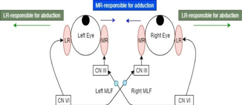
INO results from damage to the medial longitudinal fasciculus (MLF), a highly myelinated interneuron tract that connects the ipsilateral CN III to the contralateral CN VI. Therefore, MLF is responsible for coordinating conjugate lateral gaze.
A lesion in this vital interneuron breaks the connection between an ipsilateral abducting extra-ocular movement and a contralateral conjugate adduction. Damage to the MLF results in an ipsilateral adduction deficit with contralateral horizontal abduction saccades. However, convergence is intact in patients with INO, which is an important distinguishing factor from mimicking pathologies such as myasthenia gravis or a CN III palsy. INO can result from a variety of pathophysiological processes including demyelination, ischemia, inflammation and can present bilaterally or unilaterally. Ischemia is the most common pathophysiologic etiology of INO, whereas multiple sclerosis-associated demyelinating lesion is the most common cause of bilateral INO [3]. INO after cardiac catheterization is a rare phenomenon; one study estimated the frequency to be 1/1,000 – 1/6,465 cases per year and that it may be under-recognized due to spontaneous recovery of the condition [4]. The underlying pathophysiology is thought to be microembolization. The MLF is at increased susceptibility to micro emboli in comparison to surrounding arteries due to receiving its vascular supply from penetrating arteries originating from the superior one centimeter of the basilar artery [4].
Treatment of INO varies based on the underlying pathophysiology. Patients developing INO following an ischemic cerebrovascular accident are recommended to be hospitalized for full neurological assessment to rule out coincident focal neurological defects which worsen the prognosis of INO. Though not thoroughly studied, prognosis of ischemic INO is considered favorable. Many patients who develop INO, such as our patient, see their condition self-resolve and do not have any long- term visual deficits from the event.
Ocular sequelae following cardiac catheterization are rare; however, examples of vision-threatening ocular pathology associated with this common cardiac procedure have been reported. INO, which is usually benign, is thought to be due to microembolization of the MLF and the patient typically becomes asymptomatic a few months after the procedure. Specifically, Eggenberger et al. performed a retrospective analysis of isolated INO following cardiac catheterization and found that diplopia resolved with a mean duration of 82 days [4]. All patients undergoing diagnostic or therapeutic cardiac catheterization carry a small risk of ocular complications. Early recognition is important in order to optimize patient care in these rare situations.
References
- Benjamin EJ, Blaha, MJ, Chiuve SE, Cushman M, Das SR, et al. Heart Disease and Stroke Statistics-2017 Update: A Report From the American Heart Association. Circulation. 2017; 135: 146-603.
- Al-Hijji M A, Lennon R J, Gulati R, El Sabbagh A, Park J Y, et al. Safety and Risk of Major Complications With Diagnostic Cardiac Catheterization. Circ Cardiovasc Interv. 2019; 12: e007791.
- Eggenberger E R, J H Pula, Chapter 24 - Neuro-ophthalmology in Medicine, in Aminoff’s Neurology and General Medicine (Fifth Edition), Aminoff M J, Josephson S A. Editors. Academic Press. 2014: 479-502.
- Eggenberger ER, Desai NP, Kaufman DI, Pless M. Internuclear ophthalmoplegia after coronary artery catheterization and percutaneous transluminal coronary balloon angioplasty. Journal Of Neuro-Ophthalmology: The Official Journal Of The North American Neuro-Ophthalmology Society. 2000; 20: 123-126.
- Al-Hijji, MA, Lennon RJ, Gulati R, El Sabbagh A, Park JY, et al. Safety and Risk of Major Complications With Diagnostic Cardiac Catheterization. Circulation: Cardiovascular Interventions. 2019; 12: e007791.
MedDocs Publishers
We always work towards offering the best to you. For any queries, please feel free to get in touch with us. Also you may post your valuable feedback after reading our journals, ebooks and after visiting our conferences.


