Annals of Cardiology and Vascular Medicine
HOME /JOURNALS/Annals of Cardiology and Vascular Medicine- Review Article
- |
- Open Access
- |
- ISSN: 2639-4383
Eye diseases associated with systemic hypertension: What a non-ophthalmologist should know?
- Rafael Cicconi Arantes*;
- Department of Ophthalmology, Clínica de Olhos Leitão Guerra, Rua Catharina Paraguaçú, 40150-200 Salvador - BA, Brazil
- Juliana Abreu Rio;
- Department of Ophthalmology, Clínica de Olhos Leitão Guerra, Rua Catharina Paraguaçú, 40150-200 Salvador - BA, Brazil
- Lana Martins Menezes;
- Department of Ophthalmology, Clínica de Olhos Leitão Guerra, Rua Catharina Paraguaçú, 40150-200 Salvador - BA, Brazil
- Ricardo Luz Leitao Guerra
- Department of Ophthalmology, Clínica de Olhos Leitão Guerra, Rua Catharina Paraguaçú, 40150-200 Salvador - BA, Brazil

| Received | : | Mar 30, 2020 |
| Accepted | : | Apr 24, 2020 |
| Published Online | : | Apr 28, 2020 |
| Journal | : | Annals of Cardiology and Vascular Medicine |
| Publisher | : | MedDocs Publishers LLC |
| Online edition | : | http://meddocsonline.org |
Cite this article: Arantes RC, Rio JA, Menezes LM, Guerra RLL. Eye diseases associated with systemic hypertension: What a non-ophthalmologist should know?. Ann Cardiol Vasc Med. 2020: 3(1); 1016.
Background
Systemic Hypertension (SH) or elevated blood pressure is a very common medical condition and it’s complications have intimal relation with cardiovascular diseases, being one of the major causes of premature death worldwide [1]. An estimated 1.13 billion people worldwide have SH and fewer than 1 in 5 people with SH have it under control [2].
SH significantly increases the risks of heart, brain, kidney and other diseases, leading to chronic conditions that make a huge impact in all heath systems and economy around the world.
The eye is considered a target organ of SH [3]. It can be affected in different ways, but the repercussion is related with vascular alterations, from ischemia to bleeding, with different presentations.
The aim of this article is to present the most common ocular complications related to SH.
Hypertensive retinopathy
The most common ocular affection from SH is Hypertensive Retinopathy (HR), which is an important sign of uncontrolled blood pressure. It occurs mainly because of the high pressure on the ocular circulation which has some peculiarities [4]. Ocular circulation is very rich and complex. It has the highest blood flow per volume in the body [4].
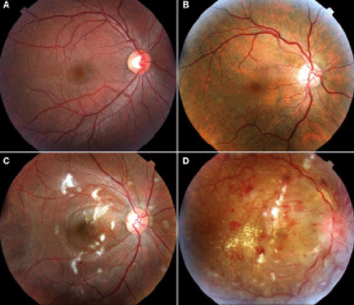
Many classifications were proposed since the HR were described. Some authors modified classic descriptions and related the retinopathy with its systemic associations. A good and practical example was described by Wong and Mitchell [5] (Table 1). It is crucial to understand that the retinopathy findings have close relation to systemic complications such as stroke, coronary heart disease and death.
Retinal vein occlusions
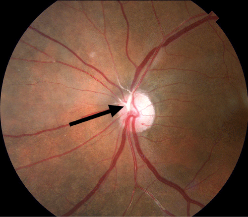
Another important ocular pathology related to the SH is the Retinal Vein Occlusion (RVO). It can occur in the central vein or in some branch of it. In the retinal arteriovenous crossings sites the vein and the artery share a common adventitious sheath allowing venular compression due to arterial sclerosis leading to branch occlusion and thrombosis [6]. It also occurs at the level of the crivous lamina where the central retinal artery and the central retinal vein share a narrow opening.
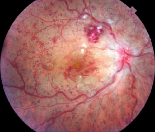
The vein occlusion leads to a blood stasis and consequently hemorrhage and exudation in the retinal tissue. As the occlusion occurs in the central vein, usually the patient complains about unilateral sudden and painless vision loss (mild or severe) and blurred vision. Pupil examination can present relative afferent pupillary defect.
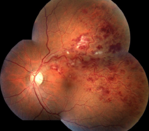
Retina examination shows venous tortuosity and dilatation, and, dot/blot and flame-shaped hemorrhages in all quadrants. Optic disc swelling and macular edema can be found. Cotton-wool patches, particularly in hypertensive patients, may be present. Transient retinal vessel wall sheathing can occur [7,6].
An occlusion can occur in a branch, and all of the findings are similar to central vein occlusion, but they are confined to the branch’s area. Clinically, the patient presents visual field defect (blurred generally) corresponding to the area of the branch with inverted correspondence (when the lesion is superior, inferior visual field is compromised, when is temporal, nasal visual field is compromised).
RVO are no urgent situations however the patients must be referred to an ophthalmologist in a short interval (less than a month) to avoid complications such as permanent vision loss and glaucoma. Timing is crucial to have the best outcome, although the visual impairment is directly related to particularities of the prime event and treatment response [8].
Retinal artery occlusions
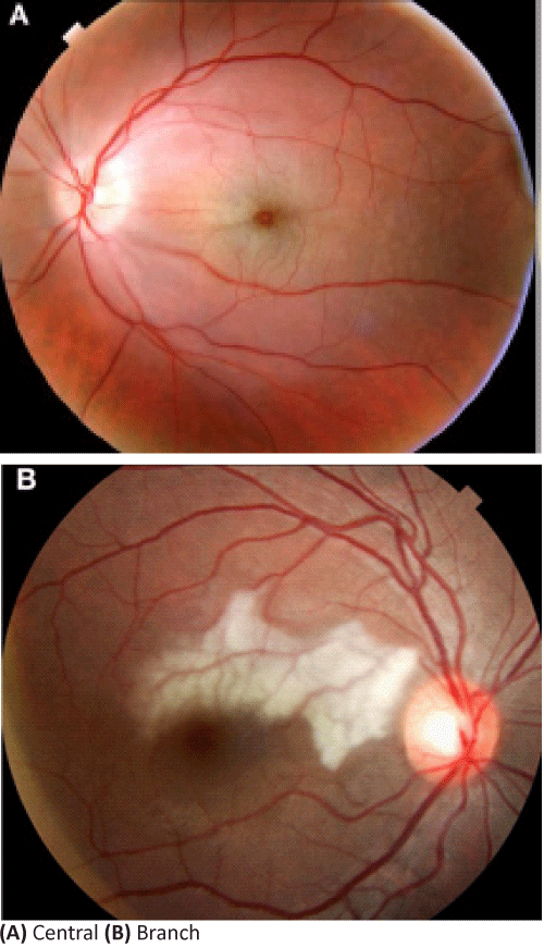
Occlusions can occur in retinal arteries, leading to central retinal artery occlusion (RAO) or branch RAO. In both cases an embolus/thrombus or a spasm leads to artery obstruction causing blood flow reduction. Retinal tissue ischemia causes severe visual loss in the non-irrigated area.
At the early onset, the diagnosis is prompted by the sudden onset of visual acuity loss and a corresponding visual field defect [9] – total when involves the central artery itself or partial when involves a branch. Usually the visual defect is more prominent than when compared to a venular occlusion, and a relative afferent pupillary defect is remarkable.
Retina examination in central RAO reveals an important diffuse retinal whitening (due to the ischemia) contrasting with the macular area (cherry-red spot sign). Eventually a plaque inside the obstructed vessel (cholesterol – Hollenhorst plaque; or calcific) can be found. The arterial branches are thinned and veins may be normal or thinned. Retinal artery occlusion is an eye emergency and the patient should be referred to the nearest stroke center for further immediate management due to an important association with acute ischemic stroke [10].
Embolism is the most common cause of arterial retinal occlusions, mainly due to atherosclerotic plaques in carotid, and the heart is another important source of thrombi [11].
Sometimes patients can experience transient visual loss (amaurosis fugax) due to a thrombotic ischemic event involving retinal arteries that resolves in a short period of time. It should be interpreted as a transient ischemic attack and the patient should be referred for investigation [12].
Despite a prompt identification and management attempts, the visual outcome of the retinal artery occlusions that affects the macula are usually poor.
Retinal arterial macroaneurysm
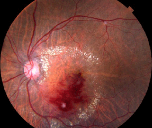
A retinal arterial macroaneurysm (RAM) is an acquired focal aneurysmal dilation of a retinal arteriole, usually within the first 3 orders of the retinal arterial system [13]. They are strongly associated with hypertension [14]. It is equivalent to a central nervous system aneurysm with similar complications and course. A RAM is uncommon but can have remarkable visual impairment.
The thickening of the arteriolar vessel wall is similar to arteriosclerotic changes elsewhere, resulting in focal areas of ischemia, remodeling of the greater intimal collagen, and, finally, vessel diameter dilation. Breaks within the arteriolar wall also result in a fusiform dilation, increasing the risk of exudation and rupture [15]. It can be an incidental finding on a routine examination in an asymptomatic person or it can be a symptomatic RAM presenting insidious or abrupt vision loss due to exudation and/or hemorrhage.
Depending on the stage of the symptoms and the visual acuity a macroaneurysm can be managed with observation, laser or even surgery (in cases of rupture and hemorrhage). Most of the cases have a good visual outcome when managed correctly and within optimal time.
Conclusion
The current review demonstrates several ocular complications due to hypertension and the potential visual impairment they can cause. The eye have the particularity that it is the only target organ in which its circulation can be seen without an invasive exam and that’s why fundus examination is an important resource to determine chronic and acute lesion of target organs.
It is important for the professionals that manage hypertension to know how ocular complications are related with the disease and its consequences. The ophthalmologist is an important allied and can provide valuable information for disease control.
Prevention of ocular complications are done with correct clinical management and referral for ophthalmic evaluation. It can avoid vision impairment or minimize damage as it occurs.
References
- Nalini M, Oranuba E, Poustchi H, et al. Causes of premature death and their associated risk factors in the Golestan Cohort Study, Iran. BMJ Open. 2018; 8.
- Bloch MJ. Worldwide prevalence of hypertension exceeds 1.3 billion. J Am Soc Hypertens. 2016; 10: 753-754.
- Chatterjee S, Chattopadhya S, Hope-Ross M, Lip P. Hypertension and the eye: changing perspectives. J Hum Hypertens. 2002; 16: 667-675.
- Flammer J, Konieczka K, Bruno RM, Virdis A, Flammer AJ, et al. The eye and the heart. Eur Heart J. 2013; 34: 1270-1278.
- Wong TY, Mitchell P. Hypertensive Retinopathy. N Engl J Med. 2004; 351: 2310-2317.
- Battaglia Parodi M, Bandello F. Branch retinal vein occlusion: Classification and treatment. Ophthalmologica. 2009; 223: 298-305.
- Hayreh SS. Classification of Central Retinal Vein Occlusion. Ophthalmology. 1983; 90: 458-474.
- Liu H, Li S, Zhang Z, Shen J. Predicting the visual acuity for retinal vein occlusion after ranibizumab therapy with an original ranking for macular microstructure. Exp Ther Med. 2018; 15: 890-896.
- Varma DD, Cugati S, Lee AW, Chen CS. A review of central retinal artery occlusion : clinical presentation and management. Eye. 2013; 27: 688-697.
- Lee J, Kim SW, Lee SC, Kwon OW, Kim YD, et al. Co-occurrence of acute retinal artery occlusion and acute ischemic stroke: Diffusion-weighted magnetic resonance imaging study. Am J Ophthalmol. 2014; 157: 1231-1238.
- Hayreh SS. Acute retinal arterial occlusive disorders. Prog Retin Eye Res. 2011; 30: 359-394.
- Kwan K, Yuen CA, Kattah JC. Update on the evaluation of transient vision loss. 2016: 297-303.
- Robertson DM. Macroaneurysms of the retinal arteries. Trans Am Acad Ophthalmol Otolaryngol. 1973; 77: OP55-67.
- Panton RW, Goldberg MF, Farber MD. Retinal arterial macroaneurysms : risk factors and natural history. 1990: 595-600.
- Streeten BW. A Histopathologic study of retinal arterial aneurysms. Am J Ophthalmol. 1978; 85: 509-513&515-518.
MedDocs Publishers
We always work towards offering the best to you. For any queries, please feel free to get in touch with us. Also you may post your valuable feedback after reading our journals, ebooks and after visiting our conferences.


