Annals of Cardiology and Vascular Medicine
HOME /JOURNALS/Annals of Cardiology and Vascular Medicine- Case Report
- |
- Open Access
- |
- ISSN: 2639-4383
Ebstein Anomaly without Fetal Hydrops but with Placentomegaly
- Aki Mabuchi*;
- Department of Obstetrics and Gynecology, Kyoto Prefectural University of Medicine, Graduate School of Medical Science, Kyoto, Japan.
- Yukiko Tanaka;
- Department of Obstetrics and Gynecology, Kyoto Prefectural University of Medicine, Graduate School of Medical Science, Kyoto, Japan.
- Miyoko Waratani;
- Department of Obstetrics and Gynecology, Kyoto Prefectural University of Medicine, Graduate School of Medical Science, Kyoto, Japan.
- Jo Kitawaki
- Department of Obstetrics and Gynecology, Kyoto Prefectural University of Medicine, Graduate School of Medical Science, Kyoto, Japan.

| Received | : | Oct 28, 2020 |
| Accepted | : | Nov 24, 2020 |
| Published Online | : | Nov 30, 2020 |
| Journal | : | Annals of Cardiology and Vascular Medicine |
| Publisher | : | MedDocs Publishers LLC |
| Online edition | : | http://meddocsonline.org |
Cite this article: Mabuchi A, Tanaka Y, Waratani M, Kitawki J. Ebstein Anomaly without Fetal Hydrops but with Placentomegaly. Ann Cardiol Vasc Med. 2020: 3(1); 1033.
Abstract
This report describes a case of Ebstein anomaly with severe tricuspid regurgitation, severe pulmonary regurgitation, and reversal of flow in the ductus arteriosus. The case was diagnosed as an Ebstein anomaly with circular shunt. Although fetal cardiomegaly had progressed and various indicators of severity of Ebstein anomaly were worsening with advancing gestational age, fetal hydrops was not detected. The pregnant woman was hospitalized from 34 weeks of gestation, and the status of the fetus was strictly monitored. At this time, transabdominal echocardiography showed that the placenta was bright and thickened to 6.4 cm, but fetal hydrops was not observed. At 36 weeks of gestation, emergent cesarean section was performed with pediatricians and pediatric cardiovascular surgeons present due to the non-reassuring fetal status. The neonate underwent aggressive resuscitation after birth, but the neonate died at 37 min after birth. As the neonate did not have fetal pleural effusion, ascites, or skin edema, the condition was not diagnosed as fetal hydrops. However, lung hypoplasia was likely seen with the progression of cardiac enlargement, and placentomegaly was also found. The placental weight after birth was 881 g, which was large than normal for the corresponding gestational age. Histologically, the presence of numerous immature villi suggested that the prenatal fetal condition was similar to fetal hydrops. Therefore, ultrasonography of the placenta should be performed to monitor diseases that might develop into fetal hydrops.
Case report
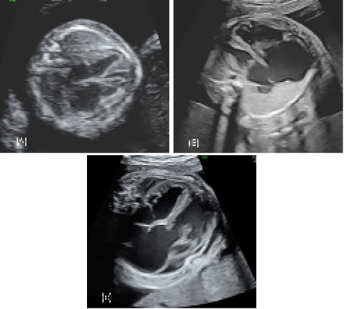
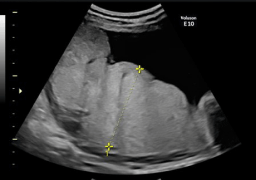
A 29-year-old healthy woman, gravida 2, with a history of early miscarriage, para 0, was referred to our hospital at 18 weeks of gestation for fetal cardiomegaly. At this time, the Cardiothoracic Area Ratio (CTAR) was 0.44, and severe Tricuspid Regurgitation (TR), severe Pulmonary Regurgitation (PR), and end-diastolic Doppler flow absence in the umbilical artery were found. Fetal hydrops was not observed. At 21 weeks, fetal cardiomegaly had progressed, showing a CTAR of 0.47. The septal and mural leaflets were displaced into the inlet component of the right ventricle, and Ebstein anomaly was diagnosed. Severe TR, continuous PR, and reversal of flow in the ductus arteriosus caused a Circular Shunt (CS), where blood was flowing from the aorta back to the pulmonary artery. The SickKids score, a predictive indicator for prognosis of Ebstein anomaly, was 8. The diameter of the ductus arteriosus was 2.3 mm. At 25 weeks of gestation, the CTAR was 0.54, the diameter of the ductus arteriosus had increased to 4.9 mm, and the backflow had increased. The Celermajer index, which predicts life prognosis based on the area ratio between the right atrium and the atrialized right ventricle in the four-chamber view, was 0.57 (grade 2); the Tei index, which evaluates cardiac function, was 0.49; and the SickKids score remained at 8. The placenta was thickened, with high echo brightness on a transabdominal echocardiogram (Figure 1), but no fetal pleural effusion, ascites, or skin edema was observed. The patient was therefore judged as not having fetal hydrops. At 34 weeks of gestation, the CTAR was 0.62; however, the fetus was growing and had an estimated body weight of 1,922 g. The Celermajer index, Tei index, and SickKids score were 0.7, 0.35, and 8, respectively; the umbilical artery had an antegrade blood flow, and the fetal status was not deteriorating. The pregnant woman was hospitalized at this time, and the fetal status was evaluated daily by cardiotocography and frequently by ultrasonography. We suspected that a ventilator and prostaglandin E1 may be required immediately after birth, and surgery was deemed necessary if the fetal status remained poor owing to the presence of a CS, despite the use of a ventilator. An elective cesarean section was scheduled at 37 weeks of gestation with pediatricians and pediatric cardiovascular surgeons present due to the non-reassuring fetal status. However, an end-diastolic Doppler flow was absent in the umbilical artery at 36 weeks and 3 days, and an emergent cesarean section was performed. Before delivery, the fetus had a CTAR of 0.65, severe TR with a flow velocity of 242 cm/s, arterial tubule diameter of 4.6 mm, retrograde flow velocity of 160.5 cm/s, Celermajer index of 0.78, Tei index of 0.72, and SickKids score of 10. At this point, pericardial effusion was observed around the heart. The neonatal birth weight was 1,878 g. The Apgar score was 1 at 1 min and 1 at 5 min. A main pulmonary artery ligation was planned immediately, but the neonate died at 37 min after birth. The placental weight after birth was 881 g, which was greater than the normal weight for the corresponding gestational age, and histologically, there were many immature villi. The mother was discharged at 5 days after the cesarean section. She provided informed consent at the time of her first visit to our hospital for the publication of this report.
Discussion
Ebstein anomaly is a rare congenital heart disease characterized by downward displacement of the tricuspid valve into the right ventricle and accounts for 0.3% of congenital heart disease cases [1-3]. The perinatal mortality rate for Ebstein anomaly is as high as 45%–80% [4-6]. The predictors of mortality include diagnosis at < 32 gestational weeks, pericardial effusion, and PR at the time of diagnosis [5]. In addition, fetal hydrops can develop when Ebstein anomaly is complicated by a CS, the prognosis for which is poor [7,8].
In the present case, fetal hydrops was not observed, but early neonatal death occurred. Placental weight was heavier than normal for 36 weeks, and placental pathology showed many immature and edematous villi. It is speculated that the fetus was already in severe circulatory dynamics in utero, having symptoms similar to those of fetal hydrops. Fetal hydrops is diagnosed when two of the following conditions are detected on ultrasonography: fetal effusion, ascites, pericardial effusion, and skin edema [9]. It is also reported to be caused by increased interstitial fluid associated with increased capillary hydrostatic pressure, decreased oncotic pressure, increased capillary permeability, and impaired lymph return [9,10]. In this case, pericardial effusion was retained, but no other findings were observed; hence, fetal hydrops was not diagnosed. However, a CS had reduced the blood volume throughout the body; thus, the amount of umbilical artery blood sent to the placenta likely decreased, the villous tissue became hypoxic as suggested by the number of immature villi, the permeability of the villous blood vessels increased, and interstitial fluid accumulated in the intervillous space. Although fetal hydrops was not diagnosed, placental thickening and high echo brightness were observed on the echocardiogram, suggesting an increase in placental weight and placental interstitial fluid.
In this case, the postnatal fetal status was similar to that in fetal hydrops, although no evidence of fetal hydrops was found using echocardiography. However, placental hyperplasia was present. Ultrasonography of the placenta should be performed to monitor diseases that might develop into fetal hydrops.
References
- Hoffman JIE, Kaplan S. The Incidence of Congenital Heart Disease. J Am Coll Cardiol. 2002; 39: 1890-900.
- Hoffman JIE, Kaplan S, Liberthson RR. Prevalence of Congenital Heart Disease. Am Heart J. 2004; 147: 425-439.
- Lang D, Oberhoffer R, Cook A, Sharland G, Allan L, et al. Pathologic Spectrum of Malformations of the Tricuspid Valve in Prenatal and Neonatal Life. J Am Coll Cardiol. 1991; 17: 1161-1167.
- Andrews RE, Tibby SM, Sharland GK, Simpson JM. Prediction of Outcome of Tricuspid Valve Malformations Diagnosed during Fetal Life. Am J Cardiol. 2008; 101: 1046-1050.
- Freud LR, Escobar-Diaz MC, Kalish BT, Komarlu R, Puchalski MD, et al. Outcomes and Predictors of Perinatal Mortality in Fetuses with Ebstein Anomaly or Tricuspid Valve Dysplasia in the Current Era: A Multicenter Study. Circulation. 2015; 132: 481-489.
- Celermajer DS, Bull C, Till JA, Cullen S, Vassillikos VP, et al. Ebstein’s Anomaly: Presentation and Outcome from Fetus to Adult. J Am Coll Cardiol. 1994; 23: 170-176.
- Wald RM, Adatia I, Van Arsdell GS, Hornberger LK. Relation of Limiting Ductal Patency to Survival in Neonatal Ebstein’s Anomaly. Am J Cardiol. 2005; 96: 851-856.
- Wertaschnigg D, Manlhiot C, Jaeggi M, Seed M, Dragulescu A, et al. Contemporary Outcomes and Factors Associated with Mortality after a Fetal or Neonatal Diagnosis of Ebstein Anomaly and Tricuspid Valve Disease. Can J Cardiol. 2016; 32: 1500-1506.
- Society for Maternal-Fetal Medicine (SMFM); Norton ME, Chauhan SP, Dashe JS. Society for Maternal-Fetal Medicine (SMFM) clinical guideline #7: Nonimmune Hydrops Fetalis. Am J Obstet Gynecol. 2015; 212: 127-139.
- Smoleniec J, James D. Predictive Value of Pleural Effusions in Fetal Hydrops. Fetal Diagn Ther. 1995; 10: 95-100.
MedDocs Publishers
We always work towards offering the best to you. For any queries, please feel free to get in touch with us. Also you may post your valuable feedback after reading our journals, ebooks and after visiting our conferences.


