Annals of Cardiology and Vascular Medicine
HOME /JOURNALS/Annals of Cardiology and Vascular Medicine- Case Report
- |
- Open Access
- |
- ISSN: 2639-4383
Diffuse Alveolar Hemorrhage as an Initial Presentation of Vasculitis: COVID-19 Induced
- Badawaki H;
- Nephrology department, Lebanese University, Lebanon.
- Tekriti Z;
- Hemato-oncology department, Lebanese University, Lebanon.
- Najem R;
- Nephrology department, Lebanese Hospital Geitawi University Medical center, Lebanon.
- Chaddad R*
- Cardiology department, Lebanese University, Lebanon.

| Received | : | Dec 01, 2021 |
| Accepted | : | Dec 24, 2021 |
| Published Online | : | Dec 27, 2021 |
| Journal | : | Annals of Cardiology and Vascular Medicine |
| Publisher | : | MedDocs Publishers LLC |
| Online edition | : | http://meddocsonline.org |
Cite this article: Badawaki H, Tekriti Z, Najem R, Chaddad R. Diffuse Alveolar Hemorrhage as an Initial Presentation of Vasculitis: COVID-19 Induced. Ann Cardiol Vasc Med. 2021; 4(2): 1056
Keywords: Diffuse alveolar hemorrhage; Covid-19; Vasculitis; Hemoptysis; Respiratory failure.
Abstract
COVID-19 is a disease caused by SARS-CoV-2 that can trigger severe respiratory tract infection. Among COVID-19 symptoms; fever, cough and fatigue are the most commonly reported. Hemoptysis is a reported symptom that could reflect diffuse alveolar hemorrhage. The main reason for hospitalization in COVID-19 infection is the onset of acute hypoxemic respiratory failure due to viral pneumonia, pulmonary embolism or other complications.
This report highlights the case of 44-year-old male who developed alveolar hemorrhage three days after covid-19 infection associated with new onset vasculitis. The patient progressively recovered with steroids and plasmapheresis.
Introduction
Coronavirus Disease 19 (COVID-19) is a respiratory virus that spread from human to human, transmitted via droplets or direct contact with mean incubation period of 6.4 days [1]. Symptoms of COVID-19 are variable and they can range from asymptomatic to Severe Acute Respiratory Syndrome Coronavirus 2 (SARS-CoV-2) [2]. Diffuse Alveolar Hemorrhage (DAH) is a severe and potentially life-threatening disease manifestation consisting of hypoxemic respiratory failure, hemoptysis, anemia, and diffuse pulmonary infiltrates on imaging [3]. DAH is associated most commonly with pulmonary capillaritis consisting of an interstitial neutrophilic predominant infiltration of the alveolar and capillary walls with leukocytoclasis caused by systemic vasculitis, Anti-Glomerular Basement Membrane (GBM) disease, and classic autoimmune disease [4].
This is the case of a young adult who developed vasculitis triggered by recent COVID-19 infection manifesting by diffuse alveolar hemorrhage, anemia and hypoxia.
Case presentation
A 44-year-old male patient with chronic kidney disease on hemodialysis three times per week, hypertension and hyperlipidemia initially presented with dyspnea, hemoptysis and fever three days after positive COVID-19 PCR. On admission, the patient had mild respiratory distress associated with pallor and fatigue.
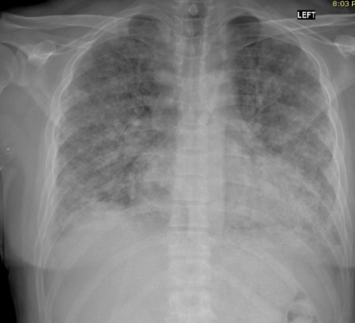
On physical examination, he had tachycardia (120 beats per min), tachypnea and his temperature was 38.5oC Fingertip oximeter measured an oxyhemoglobin saturation of 90% on 5L/min of oxygen by nasal cannula. Chest auscultation revealed bibasilar crackles. Chest X-ray showed bilateral infiltrates as shown in (Figure 1).
Initial labs showed WBC 7900, Neut 68%, Hg 5.4, Hct 16.2, MCV 85, Plat 80000/mm3, INR 1, PT 12.6, PTT 23.
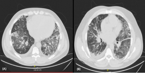
CT scanner of the chest showed diffuse reticulo-nodular densities and ground glass opacities bilaterally in the upper and lower lobes. (Figure 2).
Autoimmune workup was done and showed (ANA (1:320), Anti-cardiolipineIgG (2.7) and IgM (0.7), absent lupus anticoagulant, anti-beta2 glycoprotein (1.4), Anti-glomerular basement membrane IgG (2.43)
Bronchoscopy showed alveolar hemorrhage associated with rising RBC count in BAL and negative bacterial and fungal cultures.
The diagnosis of Diffuse Alveolar Hemorrhage (DAH) and vasculitis in a positive COVID-19 patient was made.
IV pulse steroids with several sessions of plasmapheresis were initiated. Patient’s clinical condition started to improve 1 week later with decrease in oxygen requirement and bleeding severity.
Hemoglobin improvement over 1 week was shown in Table 1after blood transfusion.
Patient was discharged home and he came again with hemoptysis and anemia so reinduction with pulse steroids was done and he started on immunosuppression with cyclophosphamide and improved over the period of 2 weeks.
Discussion
DAH is a syndrome of pulmonary hemorrhage that originates from the pulmonary microcirculation, including the alveolar capillaries, arterioles, and venules [5]. It can be categorized into four groups: Immune (vasculitis, connective tissue disease); congestive heart failure (systolic/diastolic, valvular); miscellaneous (infection, trauma, clotting disorder, cancer, drugs); idiopathic [6]. Based on a study of 34 patients with biopsy-confirmed diffuse pulmonary hemorrhage, vasculitis was the most common cause [7]. In another study of 76 patients with DAH, four parameters suggested immune-related DAH: onset of respiratory symptoms ≥ 11 days, fatigue and/or weight loss during the month prior to presentation, arthralgia or arthritis, and proteinuria ≥ 1 g/L [8].
During the COVID-19 pandemic, many cases of DAH have been reported as a rare but possible complication of COVID-19, even hemoptysis can be the first presentation of the infection [9]. Deep venous thrombosis, pulmonary embolism, large arterial thrombosis, and multiorgan venous and arterial.
Thrombosis were also reported as complications of COVID-19 infections and these manifestations have been attributed to factors such as hypoxemia, viral sepsis, immobility, and occasionally vasculitis [10]. It is unknown yet if COVID-19 associated, vasculitis is due to the direct viral invasion of the endothelium or due to the resultant immune reaction that leads to leukocytoclastic vasculitis.
In two cases reported with DAH as a complication of COVID-19 Antineutrophil Cytoplasmic Antibodies (ANCAs), antinuclear antibodies, and anti-Glomerular Basement Membrane (anti-GBM) antibodies were negative and Complement factors C3, C4 were normal excluding any autoimmune disease triggered by COVID-19 as the cause of DAH [11].
Whereas DAH was seen as the initial presentation of microscopic polyangiitis diagnosed in 77-year-old female with a recent history of recovered COVID-19 infection illustrating the possible role of COVID-19 in initiation of this autoimmune disease [12].
Our patient developed DAH while he is infected by COVID -19 and found to have Antinuclear antibody titer highly positive reflecting a possible autoimmune condition as the cause of DAH especially that the patient had a significant improvement in lung lesions with steroids and plasmapheresis.
Conclusion
This article presented an uncommon case of vasculitis, COVID-19 induced, who was successfully managed by a combined treatment of steroids, plasmapheresis and cyclophosphamide. COVID-19 virus could be a trigger for autoimmune disease in the population.
References
- Lai CC, Shih TP, Ko WC, Tang HJ, Hsueh PR. Severe Acute Respiratory Syndrome Coronavirus 2 (SARS-CoV-2) and Coronavirus Disease-2019 (COVID19): The epidemic and the challenges. Int J Antimicrob Agents. 2020; 55: 17.
- CDC COVID-19 Response Team. Severe Outcomes among Patients with Coronavirus Disease 2019 (COVID-19)-United States. MMWR Morb Mortal Wkly Rep. 2020; 69: 343-346.2.
- Collard HR, Schwarz MI. Diffuse alveolar hemorrhage. Clin Chest Med. 2004; 25: 583-592.
- Colby TV, Fukuoka J, Ewaskow SP, Helmers R, Leslie KO. Pathologic approach to pulmonary hemorrhage. Ann DiagnPathol. 2001; 5: 309-319.
- Moo Suk Park. Diffuse alveolar hemorrhage. Tuberculosis and Respiratory Diseases 2013; 74: 151-162.
- Chepuri R, Melamed K, Thomson C. Alveolar hemorrhage syndromes (diffuse alveolar hemorrhage), Decision support in medicine pulmonary medicine.
- Travis WD, Colby TV, Lombard C, Carpenter HA. A clinicopathologic study of 34 cases of diffuse pulmonary hemorrhage with lung biopsy confirmation Am J SurgPathol. 1990; 14: 1112-1125.
- Picard C, Cadranel J, Porcher R. Prigent H, Levy P, et al. Alveolar hemorrhage in the immunocompetent host: a scale for early diagnosis of an immune cause. Respiration. 2010; 80: 313-320.
- Peys E, Stevens D, Weyhaerde Y, Malfait T, Hermie L, et al. Haemoptysis as the first presentation of COVID-19: a case report. BMC Pulmonary medicine. 2020; 20: 275.
- Oxley TJ, Mocco J, Majidi S, et al. Large-vessel stroke as a presenting feature of COVID-19 in the young. N Engl J Med. 2020; 382: e60.
- Loffler C, Mahrhold J, Fogarassy P, Beyer M, Hellmich B. Two immunocompromised Patients with Diffuse Alveolar Hemorrhage as a Complication of Severe Coronavirus Disease 2019. Chest. 2020; 158: e215-e219.
- Patel R, Amrutiya V, Baghal M, Shah M, Lo A. Life-Threatening Diffuse Alveolar Hemorrhage as an Initial Presentation of Microscopic Polyangiitis: COVID-19 as a Likely Culprit. Cureus. 2021; 13: e14403.
MedDocs Publishers
We always work towards offering the best to you. For any queries, please feel free to get in touch with us. Also you may post your valuable feedback after reading our journals, ebooks and after visiting our conferences.


