Annals of Cardiology and Vascular Medicine
HOME /JOURNALS/Annals of Cardiology and Vascular Medicine- Case Report
- |
- Open Access
- |
- ISSN: 2639-4383
Delayed Management of a Myocardial Infarction Due to the Outbreak Coronavirus COVID-19
- Hery Andrianjafy*;
- Emergency Department, Groupe Hospitalier Nord Essonne Hôpital de Longjumeau, France
- Trung Hung Ta;
- Mobile Emergency and Resuscitation Service, Groupe Hospitalier Nord Essonne Hôpital de Longjumeau, France
- Giam Tran-Minh
- SOS Doctors, Chevannes, France

| Received | : | May 30, 2020 |
| Accepted | : | Jun 23, 2020 |
| Published Online | : | Jun 25, 2020 |
| Journal | : | Annals of Cardiology and Vascular Medicine |
| Publisher | : | MedDocs Publishers LLC |
| Online edition | : | http://meddocsonline.org |
Cite this article: Andrianjafy H, Trung HT, Tran-Minh G. Delayed Management of a Myocardial Infarction Due to the Outbreak Coronavirus COVID-19. Ann Cardiol Vasc Med. 2020: 3(1); 1018.
Abstract
We report a case of a patient suffering chest pain with finally myocardial infarction diagnosis. His medical care was delayed because of the COVD-19 outbreak that disrupted health care systems. We discuss the importance to maintain access to quality emergency care for patients.
Introduction
Infection with COVID-19 has spread worldwide very quickly, as noticed the World Health Organization [1]. During this dramatic pandemic disease, some countries have to face logistic and human difficulties in medical management, especially in medical care for a vital pathology. We report a case of a myocardial infarction with delay in its medical treatment.
Case presentation
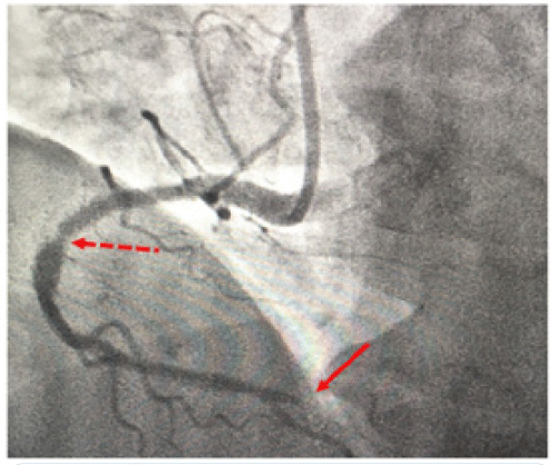
In early April 2020, a 51-year-old man with no medical history called the Emergency Medical Service (EMS) for chest pain. Given no obvious severity on telephone assessment and the COVID-19 outbreak, the man was advised to remain confined at home and recall in case of respiratory worsening or fever poorly tolerated. The next day, the patient called again EMS for increased chest pain, so an Emergency Physician (EP) went at home. The electrocardiogram (ECG) showed ischemic signs with a negative T-wave in V3V4V5, a ST elevation and a significant Q-wave in D2D3aVf, related to an inferior myocardial infarction (Figure 1). Emergency coronarography highlighted a proximal stenosis and a thrombotic distal occlusion of the right coronary artery (Figure 2).
Figure 2: Coronarygraphy: - - - Proximal stenosis of the right coronary artery ← Thrombotic distal occlusion of the right coronary artery.
Discussion
Infection with SARS-Cov-2 or COVID-19 coronavirus is a pandemic disease. All health care systems have been affected, especially in countries where the outbreak has claimed many victims [2,3]. In Italy at the height of the health crisis, pediatric EPs have noted a decrease of consultations in pediatric emergency departments (ED) for non-COVID-19 reasons. This decrease was due, firstly to the parents' fear of taking their child to an ED where there are COVID-19 cases inside, and secondly to some overwhelmed EDs that could no longer take care of other patients. This could sometimes lead to dramatic consequences [4]. In France, as of 08 April 2020, there were more than 80,000 victims of COVID-19 [5]. EMS received a considerable number of calls from people for symptoms of COVID-19, which caused congestion and risk of disruption of these services [6]. In France, in case of a phone call to the EMS for symptoms evoking an acute myocardial infarction (AMI), a medical ambulance with an EP comes to the patient’s home, and in case of a confirmed diagnosis of AMI on ECG, the patient is immediately transferred to a coronary care unit within 120 minutes as recommended in guidelines [7]. The clinical case reported here highlighted a delay in diagnosis and treatment of a AMI. This delay may have been linked to the spectacular COVID-19 outbreak, and considered a collateral damage. This delay threatened the patient’s vital and functional prognosis, considering that morbi-mortality of AMI is directly proportional to the ischaemic time duration [8].
Conclusion
The COVID-19 pandemic has disrupted health care systems, and overwhelmed medical care for life-saving emergencies. During a health crisis of this magnitude, it is essential to strengthen the EMS to ensure their role in the management of vital emergencies such as AMI, which engages the vital prognosis of victims of this pathology.
References
- Cucinotta D, Vanelli M. WHO Declares COVID-19 a Pandemic. Acta Biomed. 2020; 91: 157-160
- Frankie Tam CC, Cheung KS, Lam S, Wong A, Yung A, et al. Impact of Coronavirus Disease 2019 (COVID-19) Outbreak on ST-Segment–Elevation Myocardial Infarction Care in Hong Kong, China. Circ Cardiovasc Qual Outcomes. 2020; 13: e006631.
- Cosentino N, Assanelli E, Merlino L, Mazza M, Bartorelli AL, et al. An In-hospital Pathway for Acute Coronary Syndrome Patients during the COVID-19 Outbreak: Initial Experience under Real-world Suboptimal Conditions. Canadian Journal of Cardiology. 2020.
- Lazzerini M, Barbi E, Apicella A, Marchetti F, Cardinale F, et al. Delayed access or provision of care in Italy resulting from fear of COVID-19. 2020; 4: e10-e11.
- Santé publique France. Infection au nouveau Coronavirus (SARS-CoV-2), COVID-19, France et Monde. Mis à jour le 8 avril 2020
- Ministère des Solidarités et de la Santé. Lignes directrices pour la prise en charge par le SAMU-centre 15 des appels covid-19 en lien avec la ville. 2020.
- Ibanez B, James S, Agewall S, Antunes MJ, Bucciarelli-Ducci C, et al. 2017 ESC Guidelines for the management of acutemyocardial infarction in patients presenting with ST-segment elevation. European Heart Journal. 2018; 39: 119-177
- Scholz KH, Maier SKG, Maier LS, Lengenfelder B, Jacobshagen C, et al. Impact of treatment delay on mortality in ST-segment elevation myocardial infarction (STEMI) patients presenting with and without haemodynamic instability: results from the German prospective, multicenter FITT-STEMI trial. European Heart Journal. 2018; 39: 1065-1074.
MedDocs Publishers
We always work towards offering the best to you. For any queries, please feel free to get in touch with us. Also you may post your valuable feedback after reading our journals, ebooks and after visiting our conferences.


