Annals of Cardiology and Vascular Medicine
HOME /JOURNALS/Annals of Cardiology and Vascular Medicine- Clinical Image
- |
- Open Access
- |
- ISSN: 2639-4383
Cardiac Magnetic Resonance Imaging of Massive Left Ventricular Pseudoaneurysm
- Nijat Aliyev*;
- Medical University of Utah School of Medicine, Division of Cardiovascular Medicine, USA
- Majd Ibrahim
- Medical University of Utah School of Medicine, Division of Cardiovascular Medicine, USA

| Received | : | Jul 23, 2020 |
| Accepted | : | Aug 21, 2020 |
| Published Online | : | Aug 25, 2020 |
| Journal | : | Annals of Cardiology and Vascular Medicine |
| Publisher | : | MedDocs Publishers LLC |
| Online edition | : | http://meddocsonline.org |
Cite this article: Cite this article: Aliyev N, Ibrahim M. Cardiac Magnetic Resonance Imaging of Massive Left Ventricular Pseudoaneurysm. Ann Cardiol Vasc Med. 2020: 3(1); 1024.
Keywords: Magnetic Resonance Imaging (MRI); Imaging; Diagnostic Testing; Heart Failure; Myocardial Infarction.
Clinical image description
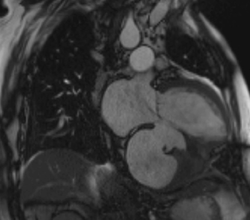
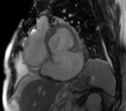
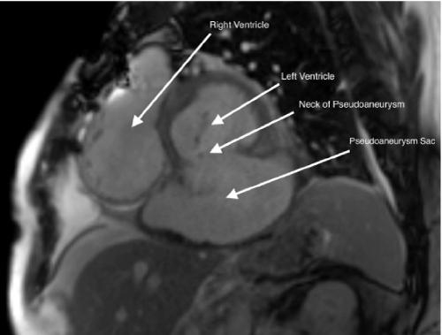
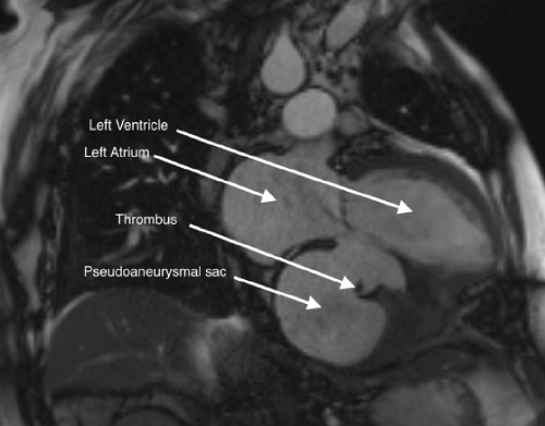
A 64-year-old man with history of coronary artery disease, end-stage renal disease presented with progressive abdominal pain. Abdominal computed tomography revealed nephrolithiasis and hemopericardium. Notably, three years prior to this presentation he had inferior ST-elevation myocardial infarction with Percutaneous Coronary Intervention (PCI) to the right coronary artery. His post PCI course was complicated by Ventricular Septal Defect (VSD) for which he underwent pericardial patch repair. During the current admission, a cardiac magnetic resonance imaging was obtained (Figure 1, Figure 3). This revealed large rupture in the basal inferior and inferoseptal segments of the myocardium with massive pseudoaneurysm and small thrombus formation (Figure 2, Figure 4, Supplementary Video – MRI Cine 2-chamber view, Video – MRI Cine short axis view). Later on, he developed heart failure and eventually underwent simultaneous heart and kidney transplantation. During post-transplant follow-up at three months, he was doing well with no reported allograft rejection.
MedDocs Publishers
We always work towards offering the best to you. For any queries, please feel free to get in touch with us. Also you may post your valuable feedback after reading our journals, ebooks and after visiting our conferences.


