- Clinical Image
- |
- Open Access
Targeting Deep Breast Lesion in a Cost Effective Way
- Teena Sleeba*;
- Consulting Radiologist, Rajagiri Hospital, Cochin Kerala, India.
- Michelle Aline Antony
- Consultant MS, Breast and Gynae-Oncology, Rajagiri Hospital, Cochin Kerala, India.

| Received | : | Aug 18, 2020 |
| Accepted | : | Sep 30, 2020 |
| Published Online | : | Oct 02, 2020 |
| Journal | : | Annals of Breast Cancer |
| Publisher | : | MedDocs Publishers LLC |
| Online edition | : | http://meddocsonline.org |
Cite this article: Sleeba T, Antony A. Targeting Deep Breast Lesion in a Cost Effective Way. Ann Breast Cancer. 2020; 3(1): 1015.
Clinical image description
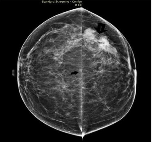
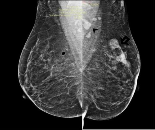
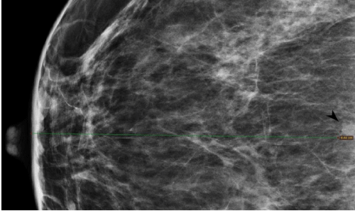
A 56 year post-menopausal lady presented with a left breast mass lesion of 3 month duration. Mammograms in CC and MLO (Figure 1,2) views revealed multifocal left breast malignancy involving the upper outer quadrant of the left breast with metastatic left axillary nodes. A subtle 4 x 4 mm nodular lesion with three specks of pleomorphic microcalcifications concerning for DCIS were also seen in the right breast (Figure 3). This finding was seen only on CC view and appeared indistinct on the RMLO view. Correlative ultrasound undertaken failed to reveal any finding.
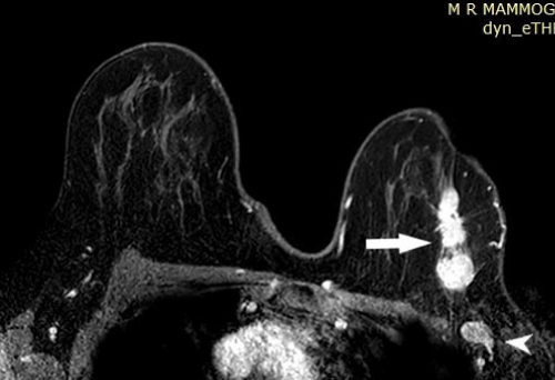
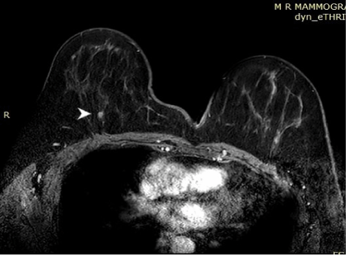
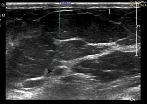
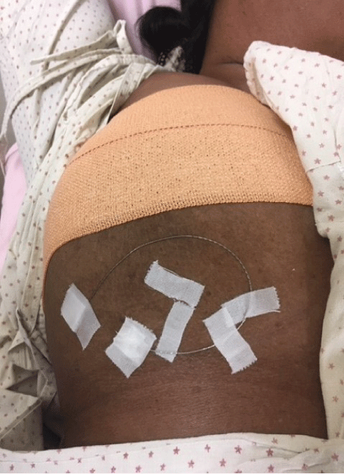
Breast MRI revealed multifocal left breast malignancy with metastatic axillary nodes (Figure 4). The right breast deeply located prepectoral nodule showed intense enhancement with true diffusion restriction (Figure 5). The lesion was seen 9 cm along the posterior nipple line in the 6’o clock position. She was taken up for a re-look USG based on the MRI location. As now the clock position of the lesion was known sonography was performed again after displacing the breast superiorly and securing it with dynaplast. A subtle 3.5 x 3 mm lesion was now seen approximately 3 cm from the skin (Figure 6). Wire localization was undertaken under USG guidance with the patient undergoing excision in this position in the theatre (Figure 7). Final histopathology revealed a grade 2 micro invasive DCIS on the right and grade 2 IDC on the left.
Stereotactically targeting extreme posterior breast lesions are tough and similarly targeting small (< 5 mm) and very deep lesions with MRI guided biopsy also has limitations. Both methods of biopsy are expensive and only limited center’s are equipped with the same.
In this case MRI helped localize this lesion following which displacing the breast significantly brought down skin-lesion distance and helped the surgeon carry out an upfront excision biopsy.
Figure 1: CC and MLO views showing multifocal left breast malignancy (thick arrow) with metastatic axillary nodes (arrow head).The right breast appears unremarkable except for a small very subtle posterior nodular lesion (small arrow).
Figure 2: CC and MLO views showing multifocal left breast malignancy (thick arrow) with metastatic axillary nodes (arrow head).The right breast appears indistinct on MLO view (small arrow).
Figure 3: A magnified view showing the right breast lesion with three small specks of pleomorphic microcalcifications (black arrowhead). This lesion is approximately 8.5cms from the nipple.
Figure 4: Dynamic post contrast enhanced breast MRI on a 3T system showing heterogeneously enhancing multifocal left breast malignancy (white arrow) with metastatic nodes (arrowhead )and intense uptake in the right deep posterior subcentimetric lesion( white arrowhead).This lesion is seen approximately 8.5 cms from the skin.
Figure 5: Dynamic post contrast enhanced breast MRI on a 3T system showing heterogeneously enhancing multifocal left breast malignancy (white arrow) with metastatic nodes (arrowhead ) and intense uptake in the right deep posterior subcentimetric lesion( white arrowhead).This lesion is seen approximately 8.5 cms from the skin.
Figure 6: Re-look USG revealed the 3.3 x 3.2 mm nodule (black arrow head). The lesion was located after superior displacement of breast and was now seen approximately 2.5 cms from the skin.
MedDocs Publishers
We always work towards offering the best to you. For any queries, please feel free to get in touch with us. Also you may post your valuable feedback after reading our journals, ebooks and after visiting our conferences.


