- Case Report
- |
- Open Access
Primary male breast leiomyosarcoma: An unusual case successfully treated by surgery and radiotherapy
- Mohammed Afif;
- Radiotherapy department, National institute of oncology, Rabat, Morocco
- Fadila Kouhen;
- Radiotherapy department, Cheikh Khalifa hospital, Casablanca, Morocco
- Hanan Elkacemi;
- Radiotherapy department, National institute of oncology, Rabat, Morocco
- Noureddine Benjaafar
- Radiotherapy department, National institute of oncology, Rabat, Morocco

| Received | : | Apr 22, 2019 |
| Accepted | : | Jun 10, 2019 |
| Published Online | : | Jun 12, 2019 |
| Journal | : | Annals of Breast Cancer |
| Publisher | : | MedDocs Publishers LLC |
| Online edition | : | http://meddocsonline.org |
Cite this article: Afif M; Kouhen F, Elkacemi H, Benjaafar N. Primary male breast leiomyosarcoma: An unusual case successfully treated by surgery and radiotherapy. Ann Breast Cancer. 2019; 2(1): 1011.
Abstract
Background: Primary sarcoma of the breast represent less than 1% of all breast malignities, and leiomyosarcoma are extremely rare. We report an unusual case of leiomyosarcoma of the breast in a Moroccan man.
Clinical case: we report a case of a 82-year-old Moroccan man, with history of larynx cancer, who present a nodule in left breast appeared six months ago. Exploration by mammography and sonography revealed a mobile and limited nodule, measuring 33x29x17mm, containing calcifications. Staging including a thoraco-abdominal CT was normal. The Patient underwent left lumpectomy without axillary dissection, histology revealed a well demarked tumor, infiltred by a proliferation with fascicular architecture, the tumor comes into contact with limits of surgical resection. Immunohistochemistry showed positivity of actin and caldesmone, and negativity for keratin, resulting in a diagnosis of leiomyosarcoma. Complementary surgery by left mastectomy followed by adjuvant radiotherapy to the chest wall was performed. 49 months after surgery, the patient is disease free.
Conclusion: Leiomyosarcoma of the breast is an extremely rare tumor that can be a diagnostic challenge to clinicians as it has no specific clinical or radiological features. It appears to have a better prognosis than other sarcomas of the breast with good outcomes after surgery.
Keywords: Leiomyosarcoma; Breast cancer; Male breast
Introduction
Sarcomas of the breast represent a rare entity with less than 1% of breast cancer [1]. Leiomyosarcoma are less common, account for under 1% of all breast sarcoma recorded in both sexes. Only 4 cases of male breast leiomyosarcomas were reported in the literature [2].
We report a case of a man followed for primary leiomyosarcoma of left breast, he was treated by mastectomy and adjuvant radiotherapy, with a good outcome after 49 months. Our case is the first case reported in Morocco.
Case presentation
An 82-year-old Moroccan man, with history of stage II squamous cell carcinoma of the larynx, treated by surgery nine years ago, was admitted to the National Oncology Institute of Rabat, for management of left breast lump.
History of this disease dates back to seven months before diagnosis, by appearance of a nodule in left breast. This mass had enlarged rapidly in the past three months, without inflammatory signs.
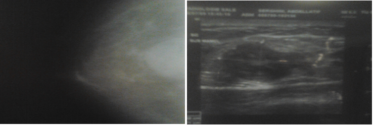
Physical examination revealed a 3 cm size mass, without a palpable axillary or supraclavicular lymphadenopathy. The controlateral breast was normal. Exploration by mammography revealed a mobile and limited nodule, with regular contours, measuring 33x29x17mm, containing calcifications. Ultrasonography confirmed suspicious malignancy (Figures 1 (a) and (b)). Staging including a thoraco-abdominal CT and bone scan was normal.
Figure 1: A&B: Preoperative mammography (a) and sonography (b), showed a limited nodule, with regular contours, measuring 33x29x17mm, containing calcifications.
The Patient underwent left lumpectomy without axillary dissection. Histo-pathological examination revealed a well demarked tumor, infiltred by a proliferation with fascicular architecture, composed of spindle-shaped cells, with eosinophil cytoplasm and irregular neucleus nucleolated sometimes monstrous. Rare atypical multinucleated cells were observed, with few mitoses and without necrosis (Figure 2). The tumor comes into contact with limits of surgical resection. Immunohistochemistry showed positivity of actin and caldesmone, and negativity for keratin and PS100. According to this pathological analysis, the tumor was diagnosed as leiomyosarcoma. A total mastectomy was performed, with no residual tumor.
Figure 2: Histophatology: spindle-shaped cells, with eosinophil cytoplasm and irregular neucleus, and few mitosis (x200).
Our case was discussed in the tumor board meeting, and adjuvant radiotherapy was indicated, a total of 50 Gy to the chest wall was delivered. No chemotherapy or hormone therapy was indicated. 49 months after surgery, the patient is disease free.
Discussion
Primary sarcomas of the breast are a rare tumors, it represent less than 1% of all breast malignancies [1]. Leiomyosarcoma are uncommon, only 30 cases was reported in the literature including 4 cases in men [2].
Origin of leiomyosarcoma of the breast is controversial. Pathologists suggest that may originate from the muscular blood vessels or from the smooth muscle of the nipple [3,4].
Diagnosis of breast primary leiomyosarcoma can be a challenge because the rarity of this entity, and of the nonspecific clinical and radiological findings. The role of the pathologist is important, and the diagnosis is mostly confirmed by the IHC [5]. Differential diagnosis includes leiomyoma, sarcomatoid carcinomas, and other soft sarcomas. Immunohistochemical staining is essential of diagnosis, leiomyosarcomas are usually positive for desmin, and actin, and are negative for S100, cytokeratins, and epithelial markers [6,7].
Optimal treatment of primary breast leiomyosarcoma is controversial due to the rarity this entity. Only a large excision with wide margins can provide good results. Axillary lymph node dissection is unnecessary because of low risk of node involvement [8]. Chemotherapy, radiotherapy and hormone therapy have not improved disease free or overall survival [1]. For our patient, surgery and adjuvant radiotherapy was sufficient to ensure control of the disease.
Leiomyosarcoma appears to have a better prognosis than other sarcomas of the breast, and the prognostic factors are not known because of the limited number of described cases. Local recurrence and lung metastasis are not rare; so the close follow-up is justified [9,10].
Discussion
Breast cancer is the commonest malignancy among women. Generally the reliability is then focused on breast cancer awareness programs to improve outcomes.
Reports have shown that 57% of cancers occur in low- and middle-income countries with 50% - 90% of the underprivileged population are deprived from access to radiation facilities [3].
In developing countries, the scarcity of breast cancer awareness programs coupled with the young age and delayed presentation, the limited resources further compounds the problem and contribute to incomplete treatments and grave outcomes [4].
The culture of the multidisciplinary approach for breast cancer management is not fully adopted in developing countries. The understanding of the complexity of Breast cancer mandates the MDT approach which provides practice guidelines with positive impact on the decisions for management plans [5].
Breast cancer staging concisely summarizes the disease status, creating a framework for assessing and planning of treatment options and predict outcomes [6]. Prognostic clinical factors such as TNM classification that remains the standard methods of staging breast cancer. Other prognostic criteria considered are tumor grade, proliferation rate, estrogen and progesterone receptor expression, human epidermal growth factor 2 (HER2) expression, and gene expression prognostic panels are added into the staging system. Additional tumor biomarkers and low Oncotype DX recurrence scores may contribute to the prognosis [7].
The extensive DNA research have contributed enormously to the role of genetics in breast cancer aiding in the promising targeted therapy [8].
Surgical options for breast cancer in developing countries are governed by limitations of the available resources for treatment coupled by the scarcity of early detection strategies and wide spread misconceptions. Reported average tumor size at the initial presentation is as more than 4 centimeters in diameter [9].
The liberal adoption of mutilating mastectomies continues to account for 80% of breast cancer surgical procedures in developing countries [10]. This plays a major role in delayed presentations as women shy away from seeking early medical advice due the limited surgical options offered.
It is proven that BCS with adequate negative margins is a notable accomplishment of modern breast cancer treatment followed by the standard adjuvant radiotherapy. It supersedes the both the physical and psychological management with mastectomy [11,12]. Radiation therapy remains the standard adjuvant treatment for breast cancer. whole-breast irradiation or accelerated partial-breast irradiation is considered based on patient selection [13].
Yet the scarcity of radiation oncology centers in the underprivileged developing world demonstrates its inability to overcome the demand of the disease burden disease burden. Worldwide [14]. This review demonstrates that in the last decades liberal mastectomies remained the standard treatment for breast cancer in developing countries. With the propagation of early detection yet sporadic breast cancer awareness programs, Breast-Conserving Surgery (BCS) has gained momentum as the preferred treatment option by both the surgeons and patients provided radiotherapy facilities is ensured. equivalent Axillary surgery is currently undergoing major revisions. It is internationally agreed that women without sentinel lymph node (SLN) metastases should be spared from Axillary Lymph Node Dissection (ALND) [15].
In western countries the successful early detection strategies succeeded in endorsing BCS followed by postoperative radiotherapy which have slowly replaced the mastectomy procedures with equal overall disease-free survival rates. The emerging oncoplastic surgery has gained popularity as it allows comfort in achieving excisions of more than 20% of the breast tissue without compromising the cosmetic outcomes [16].
However, some concerns have the documented regarding the tissue rotation of the tumor bed in the process of cosmetic contouring resulting in geographical misses in radiotherapy treatment [17].
Conclusion
Primary male breast leiomyosarcoma is an uncommon tumor with good outcomes. The treatment is based on large excision, adjuvant radiotherapy can be used if breast conserving surgery. Prognosis seems better than other breast sarcomas; however, there is a need for further cases to determine the prognostic factors.
References
- Boehm D, Keller K, Schmidt M, Cotarelo C, Kern A, et al. Primary Leiomyosarcoma of the Male Breast. World J Oncol. 2010; 1: 210-212.
- Karabulut Z, Akkaya H, Moray G. Primary Leiomyosarcoma of the Breast: A Case Report. J Breast Cancer. 2012; 15: 124-127.
- Munitiz V, Rios A, Canovas J, Ferri B, Sola J, et al. Primitive leiomyosarcoma of the breast: case report and review of the literature. Breast. 2004; 13: 72-76.
- Jayaram G, Jayalakshmi P, Yip CH. Leiomyosarcoma of the breast: Report of a case with fine needle aspiration cytologic, histologic and immune-histochemical features. Acta Cytol. 2005; 49: 656-660.
- Gupta RK, Needle aspiration cytology and immunohistologic findings in a case of leiomyosarcoma of the breast, Diagnostic Cytopathology. 2007; 35: 254-256.
- Pena J, Wapnir I. Leiomyosarcoma of the breast in a patient with a 10-year-history of cyclophosphamide exposure: A case report. Cases Journal. 2008; 1: 301.
- Lee J, Li S, Torbenson M, Liu QZ, Lind S, et al. Leiomyosarcoma of the breast: A pathologic and comparative genomic hybridization study of two cases. Cancer Genetics and Cytogenetics. 2004; 149: 53-57.
- Shinto O, Yashiro M, Yamada N, Matsuoka T, Ohira M, et al. Primary leiomyo-sarcoma of the breast: report of a case. Surg Today. 2002; 32: 716-719.
- Hussien M, Sivananthan S, Anderson N, Shiels A, Tracey N, et al. Primary leiomyosarcoma of the breast: diagnosis, management and outcome. A report of a new case and review of literature. Breast. 2001; 10: 530-534.
- Chen KT, Kuo TT, Hoffmann KD. Leiomyosarcoma of the breast: A case of long survival and late hepatic metastasis. Cancer. 1981; 47: 1883-1886.
MedDocs Publishers
We always work towards offering the best to you. For any queries, please feel free to get in touch with us. Also you may post your valuable feedback after reading our journals, ebooks and after visiting our conferences.


