- Research Article
- |
- Open Access
Ultrasonography Changes After Laser Ablation of Breast Carcinoma
- Rusy Bhalla*;
- Head of Department of Oncology, Orchid Center for Laser surgery, Mumbai India.
- Seemantini Bhalla;
- Laser Oncosurgeon, Department of Oncology, Orchid Center for Laser surgery, Mumbai India.
- Duleep Bhonsale;
- Radiologist, Department of Radiology, Orchid Center for Laser surgery, Mumbai India.
- Ashish Kapadia
- Radiologist, Department of Radiology, Orchid Center for Laser surgery, Mumbai India.

| Received | : | Jun 21, 2021 |
| Accepted | : | Jul 30, 2021 |
| Published Online | : | Aug 02, 2021 |
| Journal | : | Annals of Breast Cancer |
| Publisher | : | MedDocs Publishers LLC |
| Online edition | : | http://meddocsonline.org |
Cite this article: Bhalla R, Bhalla S, Bhonsale D, Kapadia A. Ultrasonography Changes After Laser Ablation of Breast Carcinoma. Ann Breast Cancer. 2021; 4: 1020.
Abstract
Introduction: Minimal invasive treatment for breast cancer has been introduced under various technologies. Chief amongst them are ablation techniques like Laser, Radiofrequency, Micro-ablation and Cryo-ablation.
The advantages of Laser Ablation are reduced procedure time and real time visualization of tumour.
Material and procedure: 12 patients were subject to laser ablation of tumour between 2008 and 2021. All patients were unwilling to undergo formal mastectomy and were in Stage 3 by Manchester classification. The patients were scanned by sonography and histopathological diagnosis was obtained by Tru-Cut biopsy. All patients were in BIRADS 4 and 5 category.
The procedure was performed under sonography control with 60W diode laser. The outer extent of ablation was taken to be 0.5-1.5 cms outside the visible margin of the tumor. The Lymph nodes were localized with help of Pet Scan/Sonography and were similarly laserised.
Results: The sonography images were obtained immediately and at 2-3month intervals. The findings were corroborated by PET scans taken at 3 monthly intervals for 1 year.
Conclusions: Laser Ablation is a viable procedure for necrosing the Breast cancer. It has the advantage of no mutilation or scarring. It can be repeated if there is any recurrence or new occurrence. Radiological changes after Laser Ablation are very predictable and sonologists need to be aware of the findings in view of increasing acceptability of Laser Ablation in treatment of Breast carcinoma in many centers around the world.
Introduction
Breast cancer is the leading cause of cancer in women in India. The breast registry shows over 100,000 cases added to the cancer patients list of breast cancer every year. The overall survival rate after standard treatment depends on the age of patient, tumour histopathology, staging of tumour, hormonal status [1].
Most common way of diagnosing Breast cancer is Sono mammography. The BIRADS criteria have been introduced to classify and pinpoint benign and Malignant Breast cancer. Sono mammography uses high frequency probe of 7.5MHZ and this has been refined further up to 13MHZ [2].
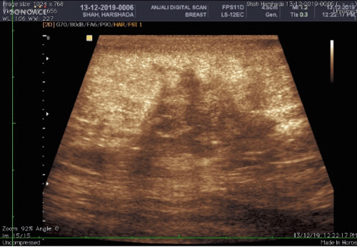
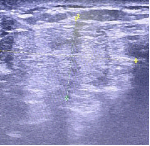
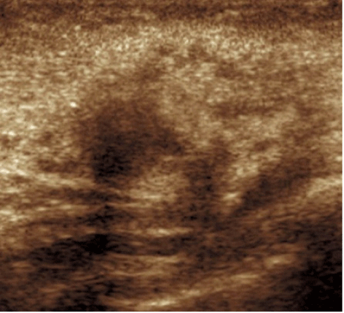
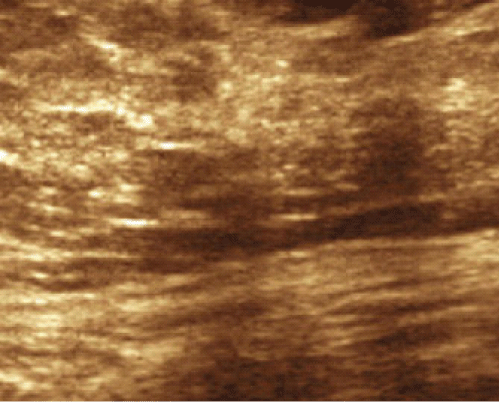
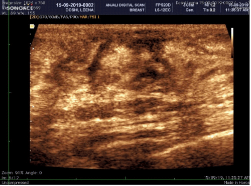
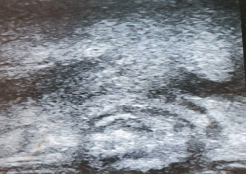
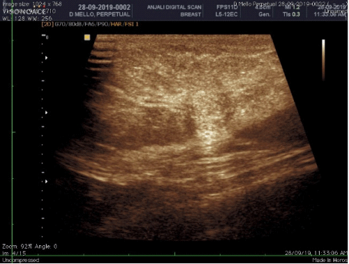
Hypoechoic. This term means “not many echoes.” These areas appear dark gray because they don’t send back a lot of sound waves. Solid masses of dense tissue are hypoechoic (Figure 2,3).
Hyperechoic. This term means “lots of echoes.” These areas bounce back many sound waves. They appear as bright gray on the ultrasound. Hyperechoic masses are not as dense as hypoechoic ones are. They may contain air, fat, or fluid (Figure 4).
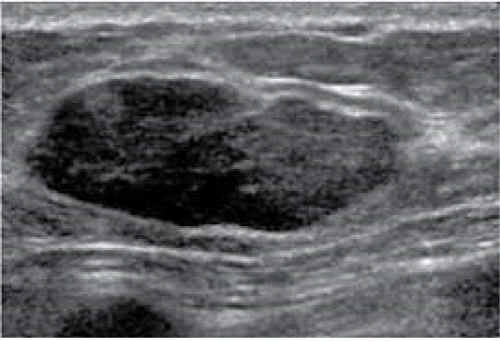
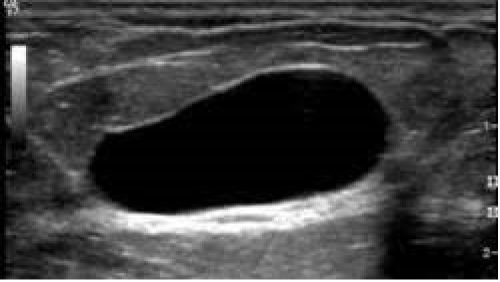
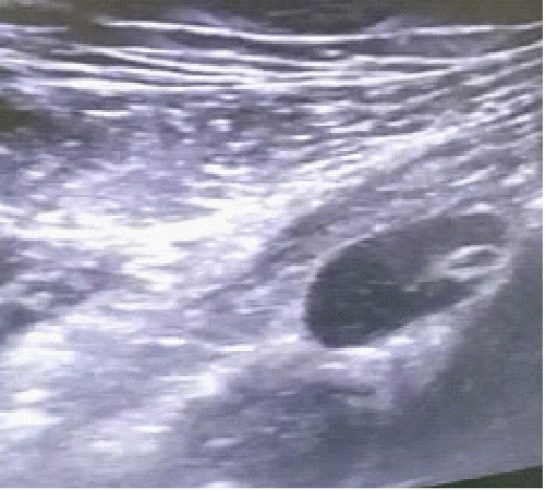
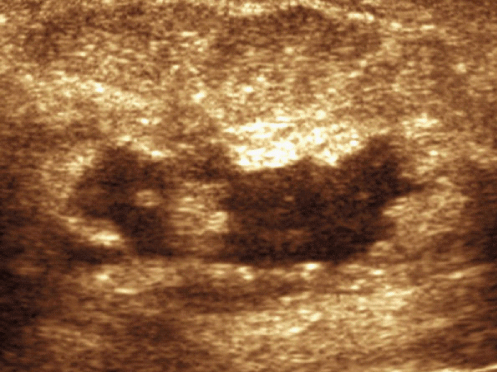
Anechoic. This term means “without echoes.” These areas appear black on ultrasound because they do not send back any sound waves. Anechoic masses are often fluid-filled and cast hyperechoic torch like area posterior to lesion (Figure 5).
The findings of different acoustics with Breast cancer is due to presence of high density areas in Breast cancers. The sound waves get absorbed in highly dense area and gives a hypoechogenic appearance on USG (Figure 2,3) [3,4,5,6].
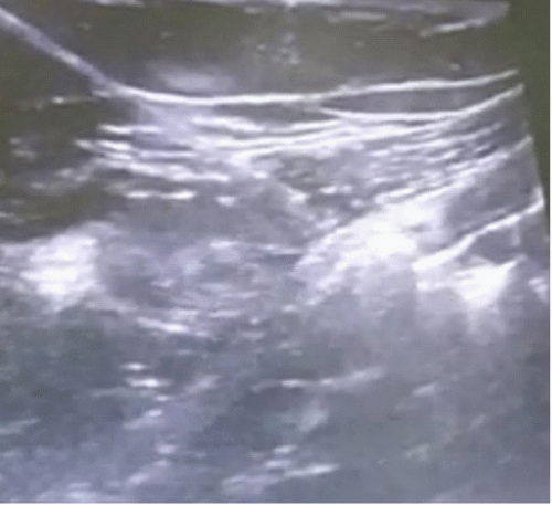
Figure 1: Shows subcutaneous hypoechoic adipose tissue and hyperechogenic breast tissue with milk glands.
Malignant lesions of Breast
The additional requirement is visualizing enlarged lymph nodes in axillary region (Figure 15). These present as enlarged oval to round structures with or without central hyperechogenic area. Absence of hilum is highly suspicious of Malignant deposits in the lymph node [7].
Normal Radiological appearance of breast
In Normal radiological appearance the normal breast tissue is hyperechogenic and layered fibro glandular tissue with hypoechogenic adipose tissue [3].
Figure 3: Classical BIRAD 5 ductal carcinoma (biopsy proven). Note: Irregular border, inhomogeneous echotexture, height more than breadth, attenuating mass.
Benign lesions of Breast
Changes during laserisation process
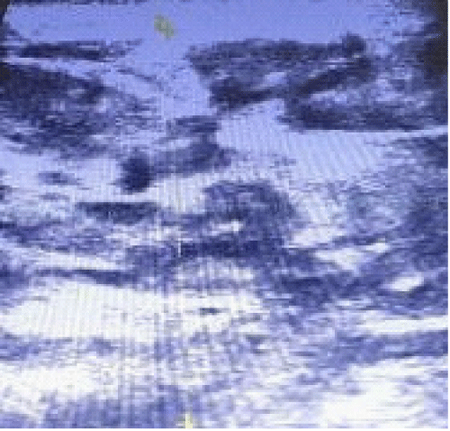
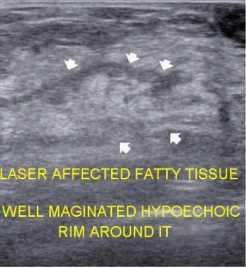
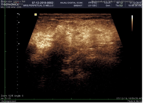
Figure 6: Hyperechogenic area with hypoechoic irregular border.The area of hyperechogenecity extends more than the dimensions of the original mass.
Changes after 2 months of laserisation
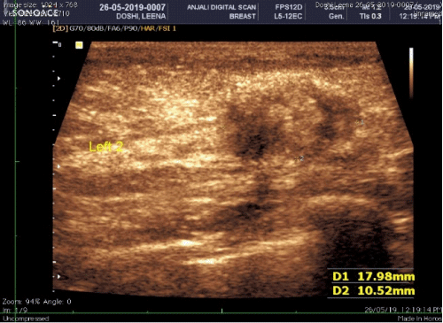
Figure 8: Cystic changes and inflammatory changes in the area of laserised mass. The anechoic area suggests cystic changes.
Changes after 6 months of laserisation
Figure 12: Inflammatory changes around rim resolving. Cystic changes change to hyperechogenic area suggestive of resolution.
Figure 14: Almost healed laserised breast lump at 6 months , Showing hyperechogenic center and inflammatory hypogenecity surrounding it.
Axillary lymph Nodes
Figure 15: Axillary Lymph node before laserisation, more hypoechoic cortex than central hyperechoic medulla.
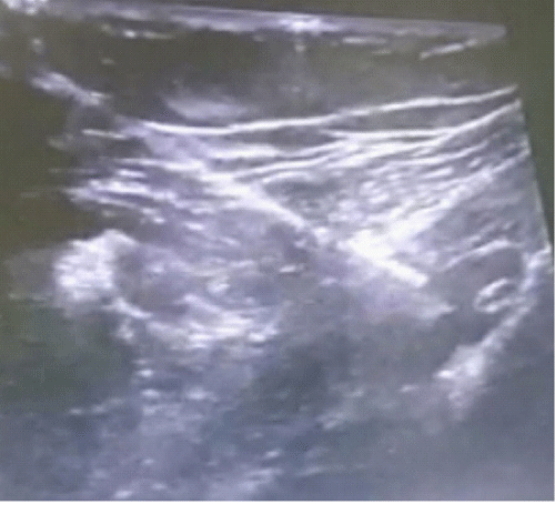
Figure 16: 18 gauze spinal needle in axillary lymph node. The laser fiber is advanced through needle in to LN for laserisation.
Changes 1 yrs after laserisation
Discussion
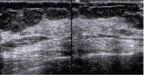
Laser Ablation of Breast cancer is fast gaining acceptability in many centers around the world [8,9, 11-14]. It is now a CE and FDA approved procedure for Breast tumours [15]. The procedure is clinically acceptable due to no mutilation of the breast. The laser procedure is in real time with acceptable necrosis of breast tumour and lymph nodes in stage 2 and 3. (Figure 6,7)
Laser works by causing a photoablation (vaporization) of tissue causing direct necrosis, Photothermal and photochemical effects-Heat denaturation of proteins and photomechanical-Thrombosis of feeding vessels due to endothelial damage and retrograde thrombosis [16].
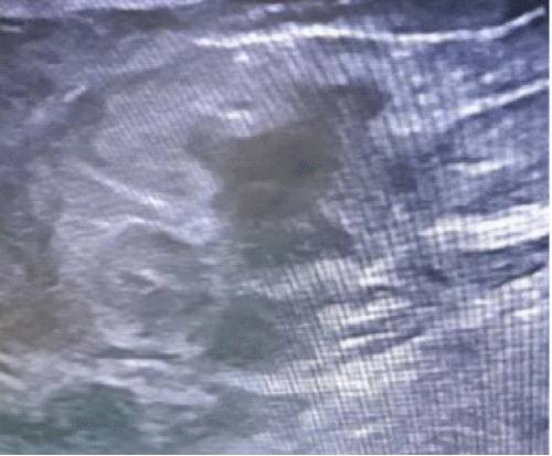
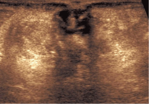
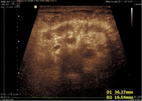
The morphological changes after laserisation is due to cell necrosis causing an interplay between hyperechogenecity and hypoechogenecity (Figure 8-14). After one year the area becomes iso-echoic or rarely hyperechogenic and may show cystic changes (Figure 19,20,21).
The initial observation of lymph node size being a criterion is no longer relevant [17,18]. Our principle is to laserised every Lymph node seen on sonography. (Figure 16,17)The visualisation of lymph nodes is guided by Previous Pet Scan and on OT-table sonography [19]. The lymph nodes show lack of vascularity and hyperechogenecity after complete laserisation. The visualized lymph node is seen after 2months (Figure 18).
After reviewing the literature, we could not find any information on the changes after laserisation.
Conclusion
Laser ablation of Breast tumours is fast gaining acceptability and sonologists need to be aware of sequential findings on follow up Sonography and PET scan of the patient.
References
- National Centre for Disease Informatics and Research: Time trends in cancer incidence rates 1982-2010. Bangalore, India, National Centre for Disease Informatics and Research, National Cancer Registry Programme, Indian Council of Medical Research, Google Scholar. 2003.
- Disha Emine, Suzana MK, Halit Y, Arben K. Comparative Accuracy of Mammography and Ultrasound in Women with Breast Symptoms According to Age and Breast Density. Bosnian journal of basic medical sciences/Udruženje basičnih mediciniskih znanosti = Association of Basic Medical Sciences. 2009; 9: 131-136.
- Alireza N, Atoosa A, Mahshid H, Morteza H. An Evaluation of Ultrasound Features of Breast Fibroadenoma. Advanced Biomedical Research. 2017; 6: 153.
- Adrada B, Wu Y, Yang W. Hyperechoic Lesions of the Breast: Radiologic-Histopathologic Correlation. American Journal of Roentgenology. 2013; 200: 5.
- Medeiros MM, Graziano L, de Souza JA, Guatelli CS, Poli MR, Yoshitake R. Hyperechoic breast lesions: Anatomopathological correlation and differential sonographic diagnosis. Radiol Bras. 2016; 49: 43-48.
- https://www.webmd.com/cancer/what-is-hypoechoic-mass
- Pinheiro DJ, Elias S, Nazário AC. Axillary lymph nodes in breast cancer patients: Sonographic evaluation. Radiol Bras. 2014; 47: 240-244.
- Jeffrey SS, Birdwell RL, Ikeda DM, et al. Radiofrequency Ablation of Breast Cancer: First Report of an Emerging Technology. Arch Surg. 1999; 134: 1064–1068.
- Schwartzberg B, Lewin J, Abdelatif O, et al. Phase 2 Open-Label Trial Investigating Percutaneous Laser Ablation for Treatment of Early-Stage Breast Cancer: MRI, Pathology, and Outcome Correlations. Ann Surg Oncol. 2018; 25: 2958-2964.
- https://www.laserfocusworld.com/biooptics/biomedicine/article/14195250/laser-ablation-technology-for-destroying-breast-tumors-gets-ce-mark-approval.
- Nori J, Gill MK, Meattini I, Paoli CD, et al. “The Evolving Role of Ultrasound Guided Percutaneous Laser Ablation in Elderly Unresectable Breast Cancer Patients: A Feasibility Pilot Study”, BioMed Research International. 2018; 2018: 9141746.
- Dowlatshahi K, Francescatti DS, Bloom KJ. Laser therapy for small breast cancers The American Journal of Surgery. 2002; 184: 359–363.
- van Esser S, Stapper G, van Diest PJ, van den Bosch MA, Klaessens JH, Mali WP, Borel Rinkes IH, van Hillegersberg R. Ultrasound-guided laser-induced thermal therapy for small palpable invasive breast carcinomas: a feasibility study. Ann Surg Oncol. 2009; 16: 2259-2263.
- Rusy Bhalla. https://meetinglibrary.asco.org/record/200975/abstract
- https://www.fda.gov/radiation-emitting-products/surgical-and-therapeutic-products/medical-lasers#p
- Lanzafame RJ. Laser/Light Applications in General Surgery. In: Nouri K. (eds) Lasers in Dermatology and Medicine. Springer, Cham. 2018.
- Koelliker SL, Chung MA, Mainiero MB, Steinhoff MM, Cady B. Axillary lymph nodes: US-guided fine-needle aspiration for initial staging of breast cancer--correlation with primary tumor size. Radiology. 2008; 246: 81-89.
- Bedi DG, Krishnamurthy R, Krishnamurthy S, Edeiken BS, Le-Petross H, et al. Cortical morphologic features of axillary lymph nodes as a predictor of metastasis in breast cancer: in vitro sonographic study. AJR Am J Roentgenol. 2008; 191: 646-652.
- Bhalla R, Bhalla S, Bhonsale D, Kapadia A. Laser Ablation of Metastatic Lymph Nodes in the Neck for Oral Carcinoma-Technique and Viability of the Procedure. Journal of Radiology and Clinical Imaging. 2021; 4: 027-035.
MedDocs Publishers
We always work towards offering the best to you. For any queries, please feel free to get in touch with us. Also you may post your valuable feedback after reading our journals, ebooks and after visiting our conferences.


