- Case Report
- |
- Open Access
- |
- ISSN: 2637-9627
An Unusual Case of Bilateral Congenital Abdominal Wall Defect in a Single Infant
- Oghogho Igbinoba;
- Department of Pediatrics, Driscoll Children’s Hospital, Corpus Christi, Texas, USA.
- Patrick Ovie Fueta*;
- Department of Pediatrics, Driscoll Children’s Hospital, Corpus Christi, Texas, USA.
- Haroon Patel
- Department of Pediatrics, Driscoll Children’s Hospital, Corpus Christi, Texas, USA.

| Received | : | Mar 07, 2022 |
| Accepted | : | Mar 23, 2022 |
| Published Online | : | Mar 28, 2022 |
| Journal | : | Annals of Pediatrics |
| Publisher | : | MedDocs Publishers LLC |
| Online edition | : | http://meddocsonline.org |
Cite this article: Igbinoba O, Fueta PO, Patel H. An Unusual Case of Bilateral Congenital Abdominal Wall Defect in a Single Infant. Ann Pediatr. 2022; 5(1): 1094.
Keyword: Abdominal wall; Gastroschisis; Left lower quadrant; Right lower quadrant.
Abbreviations: LLQ: Left Lower Quadrant; RLQ: Right Lower Quadrant; DOL: Day of Life; TPN: Total Parental Nutrition; HFOV: High-Frequency Oscillatory Ventilator; HFNC: High Flow Nasal Cannula.
Abstract
Gastroschisis is one of the most common anterior abdominal wall defects described in literature. Over the last two to three decades, the incidence of gastroschisis has trended upwards and currently stands at 1-3 per 10,000 live births (about 1,871 infants each year). The pathogenesis of gastroschisis is both poorly defined and understood. The most agreed upon etiology in the current literature is the disruption in the vascular supply of the anterior abdominal wall resulting in abdominal wall fold failure. This supports the usual single right-sided location of the defect. Rarely, a left sided defect has been described however; a bilateral musculocutaneous defect in a single infant has not been reported. We report an unusual case of congenital anterior abdominal wall defects with a right and left sided musculocutaneous defect identified in a single live infant. The defects were corrected surgically, and the patient made a full recovery. Due to the unusual presentation and anatomical locations of the defects, we cannot conclusively determine that this is a true case of bilateral gastroschisis. This emphasizes the importance of disease surveillance in order to expand our knowledge on varying presentations of congenital abdominal wall defects to formulate a more accurate hypothesis on its pathogenesis.
Introduction
Gastroschisis is one of the most common congenital ventral abdominal wall defects [1]. It is a lateral abdominal wall defect that is typically located approximately 1-2 cm to the right of the umbilicus, with evisceration of abdominal contents without a membranous covering [1,2]. Due to the lack of protective membranous covering in gastroschisis, it is associated with an increased incidence in morbidity and mortality from intestinal injury secondary to inflammation, intestinal perforation, volvulus, and atresia [1-3]. Over the last two decades, the incidence of gastroschisis has steadily risen with a current rate of 1-3 per 10,000 live births [1,4]. The exact cause of this rising statistic is unknown.
The diagnosis of gastroschisis can be made prenatally with the earliest signs being elevated alpha-fetoprotein and visualization on prenatal ultrasonography [1]. The purpose of prenatal screening and early diagnosis is to facilitate post-natal management and improve the overall prognosis [1,4].
Several unusual presentations of gastroschisis have been discussed in the literature however; they are mostly confined to left-sided defects and unusual intestinal atresia patterns. We identified one case of an infra-umbilical anterior abdominal wall defect, and one case of bilateral ventral abdominal wall defect. However, the bilateral ventral abdominal wall defect was a true musculocutaneous defect (gastroschisis) on the right and only a muscular defect on the left [5-8]. We report a case of congenital abdominal wall defect with bilateral ventral musculocutaneous openings and an intact umbilicus. This unusual presentation challenges existing definitions and the pathogenesis of congenital ventral abdominal wall defects, especially gastroschisis.
Case presentation
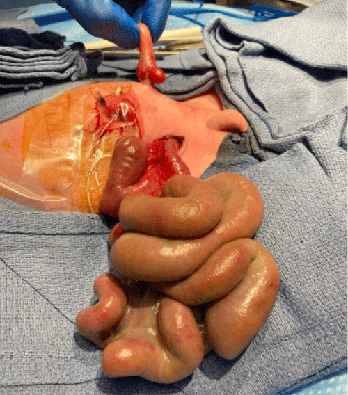
We present the case of a newborn male infant, delivered at 37 gestational weeks to a 25-year-old primigravida via induced vacuum-attended vaginal delivery for prenatal ultrasound diagnosed gastroschisis. At birth, he was found to have 2 abdominal wall defects; one in the Left Lower Quadrant (LLQ) and a second larger defect in the Right Lower Quadrant (RLQ), with evisceration of his intestines from both defects that were not covered by membranes (Figure 1).
He was immediately placed in a bowel bag and an Anderson tube was inserted with 80 mL of meconium stained fluid suctioned. He was transferred to our center on Day of Life (DOL) 0 from the birth hospital for surgical evaluation. He was subsequently intubated due to signs of respiratory distress, and he remained sedated and mechanically ventilated on presentation to our facility.
His examination findings on admission were consistent with a 1 cm anterior abdominal wall defect in the LLQ. The defect was inferior and lateral to an intact umbilicus with a single loop of intestine and his left testicle eviscerated. A second defect located between the umbilicus and the right inguinal ligament measuring approximately 2 cm was noted with a larger amount of eviscerated bowel. The intestines were dilated, edematous and inflamed. He was also noted to have bilaterally inferiorly displaced thumbs on examination. Chromosomes and microarray studies were done and results revealed no genetic abnormalities.
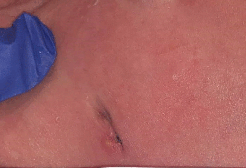
He underwent a bedside exploratory laparotomy with reduction of the LLQ contents and closure of the defect (Figure 2).
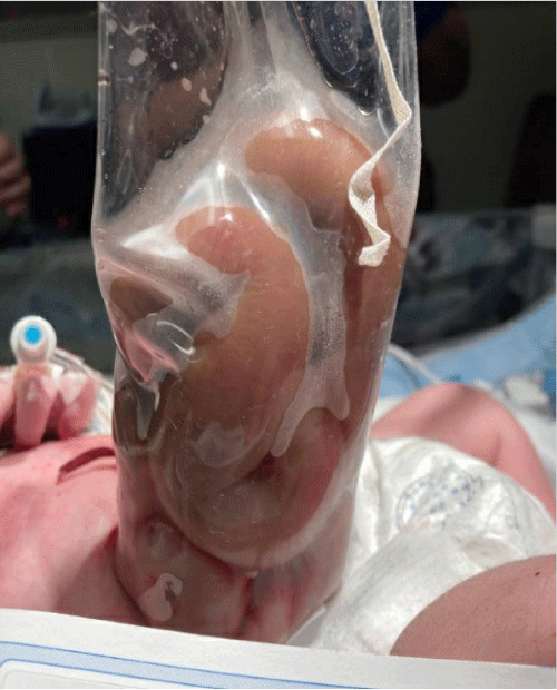
The RLQ defect was surgically enlarged to accommodate a 5.5 silo in the RLQ (Figure 3).
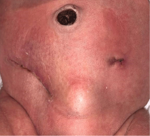
His Anderson tube was placed to low intermittent suction for gastric decompression. He tolerated the procedures without any complications and remained sedated and mechanically ventilated. He remained NPO postoperatively and Total Parenteral Nutrition (TPN) was initiated on DOL1. Low dose dopamine for bowel perfusion was administered for 2 days. From DOL2, he successfully underwent gradual staged reduction of his exposed bowel in the RLQ from the silo back into the abdominal cavity. On DOL6, he underwent a repeat exploratory laparotomy with final reduction and closure of his RLQ abdominal wall defect and placement of an incisional wound vac. Following the final closure (Figure 4), antibiotics were discontinued.
Our patient’s clinical course was complicated by worsening ventilation and oxygenation on DOL4. Capillary blood gases done at the time was significant for increasing CO2 levels, and he was switched from conventional ventilator to a High-Frequency Oscillatory Ventilator (HFOV). He subsequently improved on HFOV and was transitioned back to a conventional ventilator on DOL 8.3% hypertonic saline with chest physiotherapy were initiated to assist with his respiratory function. He was extubated to High Flow Nasal Cannula (HFNC) at 10 liters per minute and 21% FiO2 on DOL11. His surgical sites were noted to be healing well without any concern for infection. TPN was continued for nutrition until DOL13 when trophic feeds were initiated and slowly advanced while weaning the patient off HFNC to room air, which was achieved on DOL16.
Discussion
Gastroschisis is one of two well-known congenital abdominal wall defects; the other being omphalocele. Although the pathogenesis of gastroschisis is unknown, multiple hypotheses have been proposed which primarily center on the disruption in the formation of the anterior body wall during the embryonic period [5] It has been theorized to be secondary to disruption in the involutory process of the right umbilical vein, which normally occurs in the 6th week of gestation, with subsequent weakening of the body wall. This theory supports the more common presentation of gastroschisis to the right of the umbilicus. Another theory supporting the predominant finding of right-sided gastroschisis is the disruption of the right vitelline artery also resulting in weakening of the anterior abdominal wall. Although rare, a mal-closure of the left umbilical vein has been stipulated as a mechanism responsible for the development of left-sided gastroschisis. In 2007, Feldkamp et al., [9] proposed a novel theory that may be responsible for either a right or a left sided gastroschisis. Their hypothesis centers on the position of the yolk sac. It stipulates that the positioning of the yolk sack to the right, with subsequent failure of one or more folds responsible for abdominal wall closure favors gastroschisis as a right-sided abdominal wall defect. Furthermore, a slight mal-positioning of the yolk sac to the left of the midline with subsequent abdominal wall fold failure may explain the rare phenomenon of a left-sided gastroschisis.
Left-sided gastroschisis is extremely rare and as of 2013, there were only 20 reported cases in literature [5]. There have been relatively few reported cases of congenital abdominal wall defects that have deviated from the presentations described above [5,6]. In a 2002 report published by Ashburn et al., [5] it was noted that because gastroschisis has been demonstrated to be a single defect most commonly occurring to the right of the umbilicus and rarely to the left but never bilaterally in a single infant, it supports the umbilical vein theory. We did however identify one report of bilateral ventral abdominal wall defects in a single infant. The patient in this case had only muscular agenesis with an intact skin on the right side and a musculocutaneous defect on the left [7].
Our case illustrates a previously unreported bilateral musculocutaneous congenital abdominal wall defects in a single live chromosomally normal male infant. This raises the following questions; Are there alternative hypotheses yet to be described that may be responsible for the development of gastroschisis? What other hypothesis better describes a bilateral anterior abdominal wall defect lateral and inferior to an intact umbilicus?
Conclusion
Although this case was managed as a bilateral gastroschisis, we cannot definitively conclude that this is a true case of bilateral gastroschisis. If this is a case of bilateral gastroschisis, then it begs to stipulate that the underlying etiology of gastroschisis may not be entirely due to disruption of the vascular supply to the anterior abdominal wall. Also, given the increase in the incidence of gastroschisis in the last two to three decades, and the hypothesis of this incidence trend as being associated with multifactorial exposures, the importance of appropriately defining the etiology of this entity cannot be overemphasized. It is our hope that with the report of this unusual case, greater awareness is raised in the scientific community for the need of further research into congenital abdominal wall defects, possibly expanding the classification and description to abdominal wall defects rather than the traditional simple gastroschisis vs omphalocele with improved surveillance to better understand its incidence and etiology.
Declaration
Table of contents summary: This case discusses the unusual presentation of bilateral congenital abdominal was defect that deviates from currently known anatomic presentation of gastroschisis, but was successfully managed. This serves to challenge the current proposed etiologies of gastroschisis.
Author contributions: Dr. Igbinoba, conceptualized and designed the study, collected data, carried out the initial analyses, drafted the initial manuscript.
Dr. Fueta, conceptualized and designed the study, collected data, carried out the initial analyses, drafted the initial manuscript.
Dr. Patel, conceptualized and designed the study, collected data, carried out the initial analyses, critically reviewed the manuscript for important intellectual content, reviewed and revised the manuscript.
All authors approved the final manuscript as submitted and agree to be accountable for all aspects of the work.
Data availability: The data used to support the findings of this study are available on reasonable request from the corresponding author.
References
- Christison-Lagay ER, Kelleher CM, Langer JC. Neonatal abdominal wall defects. Semin Fetal Neonatal Med. 2011; 16: 164-172.
- Kastenberg ZJ, Dutta S. Ventral abdominal wall defects. NeoReviews. 2013; 14: e402-e411.
- Singh K, Kumar A. Anterior Abdominal Wall Defects, Diaphragmatic Hernia, and Other Major Congenital Malformations of the Musculoskeletal System in Barbados, 1993-2012. J Pediatr Genet. 2017; 6: 92-97.
- Montes-Tapia F, Cura-Esquivel I, Gutierrez S, Rodriguez-Balderrama I, de la OCM. Congenital lateral abdominal wall hernia. Pediatr Int. 2016; 58: 788-790.
- Ashburn DA, Pranikoff T, Turner CS. Unusual presentations of gastroschisis. Am Surg. 2002; 68: 724-727.
- Fraser N, Crabbe DC. An unusual left-sided abdominal-wall defect. Pediatr Surg Int. 2002; 18: 66-67.
- Arroyo Carrera I, Pitarch V, Garcia MJ, Barrio AR, Martinez-Frias ML. Unusual congenital abdominal wall defect and review. Am J Med Genet A. 2003; 119A: 211-213.
- Patel RV, More B, Sinha CK, Rajimawale A. Inferior gastroschisis. BMJ Case Rep. 2013: 2013.
- Feldkamp ML, Carey JC, Sadler TW. Development of gastroschisis: Review of hypotheses, a novel hypothesis, and implications for research. Am J Med Genet A. 2007; 143A: 639-652.


